Thymoma and Thymic Carcinoma: Diagnosis and Treatment
Understanding rare thymus cancers: comprehensive guide to thymoma and thymic carcinoma diagnosis, staging, and treatment options.

Understanding Thymoma and Thymic Carcinoma
The thymus is a small, butterfly-shaped gland located behind your breastbone in the upper chest, sitting in front of and above your heart. Despite its small size, the thymus plays a crucial role in your immune system by producing and maturing T-lymphocytes, which are white blood cells essential for fighting infections and protecting your body from disease. When malignant tumors develop in this vital gland, they are classified into two main types: thymoma and thymic carcinoma.
Both thymoma and thymic carcinoma are rare cancers, with approximately 400 new cases diagnosed annually in the United States. While both types originate in the thymus gland, they have distinct characteristics, growth patterns, and treatment requirements. Understanding the differences between these two conditions is essential for patients and their families when navigating diagnosis and treatment options.
Key Differences Between Thymoma and Thymic Carcinoma
Although thymoma and thymic carcinoma both develop in the thymus gland, they differ significantly in their aggressiveness and behavior. Thymoma is generally considered the less serious of the two conditions. In thymoma, tumor cells closely resemble normal, non-cancerous thymus cells. These tumors typically grow slowly and rarely spread to other parts of the body. Many patients with thymoma lead relatively normal lives with appropriate treatment.
Thymic carcinoma, by contrast, is much more aggressive. The tumor cells in thymic carcinoma look distinctly abnormal and do not resemble normal thymus cells. These cancers grow more quickly and have a higher propensity to spread to other areas of the body. By the time thymic carcinoma is discovered, it has often already metastasized beyond the thymus, making treatment more challenging.
Additionally, patients with thymoma frequently develop associated autoimmune conditions, such as myasthenia gravis and acquired pure red cell aplasia, in which the body’s immune system attacks its own healthy tissues and organs.
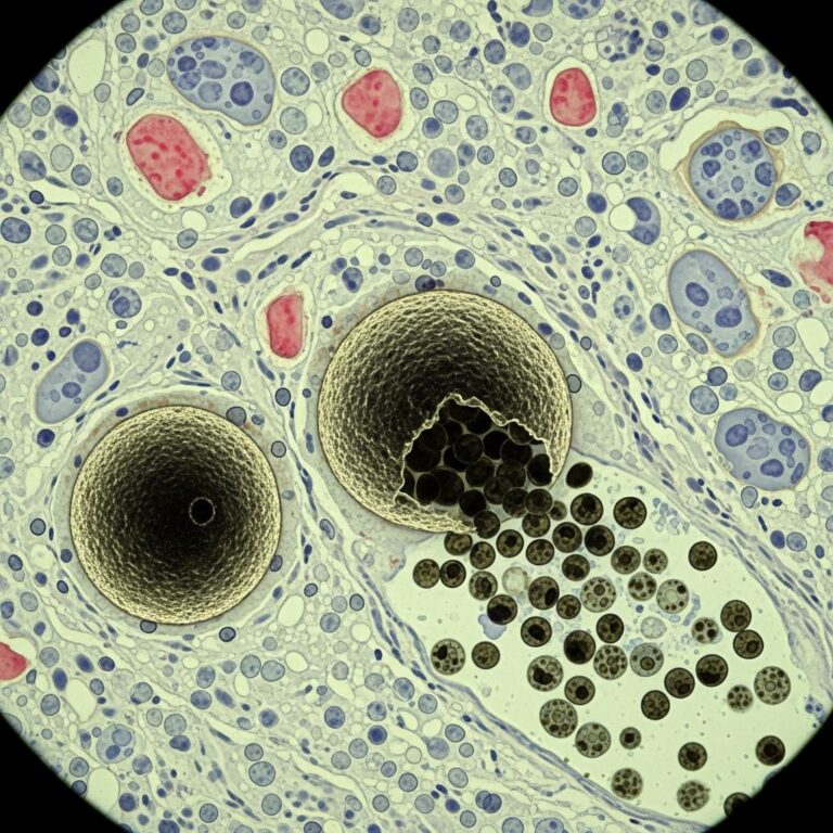
Histological Classification of Thymic Tumors
The World Health Organization (WHO) classification system categorizes thymic epithelial tumors based on their histological appearance and behavior. This classification helps clinicians predict tumor aggressiveness and guide treatment decisions.
Thymoma Types (A through B3): Type A thymomas consist of spindle or oval thymic epithelial cells with minimal lymphocytes and are considered the least aggressive. Type AB thymomas contain features of both Type A and Type B tumors. Type B1 thymomas resemble normal thymic tissue with numerous lymphocytes and few epithelial cells. Type B2 thymomas have abnormal-appearing epithelial cells with enlarged nuclei and several lymphocytes. Type B3 thymomas contain few lymphocytes and consist mostly of thymic epithelial cells that appear relatively normal.
Thymic Carcinoma (Type C): Thymic carcinomas are classified separately and are further divided into low-grade and high-grade categories based on their aggressiveness and likelihood of spreading. These tumors have clearly abnormal cells and behave more aggressively than thymomas.
Staging System for Thymic Cancers
Proper staging is critical for determining prognosis and guiding treatment decisions. The staging system helps clinicians understand how far the cancer has spread and assess the tumor burden.
| Stage | Description |
|---|---|
| Stage I | Cancer remains confined within the thymus gland without extending beyond its capsule |
| Stage II | Cancer has spread through the thymic capsule to the chest cavity lining or nearby fatty tissue |
| Stage III | Cancer has spread to structures within the chest, including the outer layer of the heart, lungs, or major blood vessels |
| Stage IVA | Cancer has spread throughout the pericardium (heart sac), lungs, or both |
| Stage IVB | Cancer has metastasized to the bloodstream or lymphatic system, indicating distant spread |
Symptoms and Warning Signs
Many patients with early-stage thymoma or thymic carcinoma experience no symptoms and may discover their condition incidentally during imaging tests performed for other reasons. However, as the tumor grows, patients may experience various symptoms depending on the tumor’s size and location. Common symptoms include chest pain or discomfort, cough, shortness of breath, and difficulty swallowing. Some patients report shoulder pain or weakness in the arms and legs. In cases where the tumor compresses nearby structures, patients may experience superior vena cava syndrome, characterized by swelling in the face, neck, and upper body.
Diagnostic Evaluation and Testing
Accurate diagnosis is essential for determining the appropriate treatment strategy. During your initial evaluation, your healthcare provider will perform a comprehensive physical examination and order several diagnostic tests to confirm the diagnosis and assess the disease extent.
Imaging Studies: CT scans and MRI imaging are the primary diagnostic tools for identifying thymic tumors. CT scans provide detailed cross-sectional images of the chest and effectively show tumor size, location, and relationship to surrounding structures. MRI offers excellent soft tissue contrast and is particularly useful for evaluating tumor invasion into adjacent organs. Positron emission tomography (PET) scans may be used to detect metastatic disease.
Biopsy: A tissue biopsy is the definitive diagnostic method for confirming cancer and determining the specific histological type. During a biopsy, a small tissue sample is obtained through needle aspiration or surgical sampling and examined under a microscope by a pathologist.
Blood Tests: Laboratory studies may help assess overall health status and monitor for any associated autoimmune conditions, particularly in patients with thymoma.
Personalized Treatment Planning
Treatment for thymoma and thymic carcinoma is highly individualized and depends on multiple factors including tumor stage, histological type, patient age, overall health status, and personal treatment goals. A multidisciplinary team approach, bringing together specialists from various fields, ensures comprehensive evaluation and optimal treatment planning.
Your treatment team may include thoracic surgeons, medical oncologists, radiation oncologists, cardiologists, pulmonologists, and supportive care specialists. Each team member contributes their expertise to develop a customized treatment strategy tailored to your specific situation.
Surgical Treatment
Surgery represents the cornerstone of treatment for most patients with thymoma and thymic carcinoma. For Stage I thymoma, surgical removal of the thymus gland (thymectomy) is the standard treatment and often provides excellent long-term outcomes. In more advanced stages, surgeons perform more extensive procedures, removing not only the thymus but also affected surrounding tissues, including lymph nodes, portions of the chest wall, portions of the lungs, or areas of the heart lining if necessary.
Advanced surgical techniques, including minimally invasive approaches, may be employed when appropriate. Surgeons at specialized centers have extensive experience managing even the most challenging cases, such as tumors that have grown around coronary artery bypass grafts. The goal of surgery is to achieve complete tumor resection while preserving as much normal tissue and organ function as possible.
Radiation Therapy
Radiation therapy uses high-energy beams to destroy cancer cells and is an important component of treatment for many patients. Radiation may be recommended in several scenarios: to shrink tumors before surgery, to destroy remaining cancer cells after surgery, or as a standalone treatment when surgery is not feasible.
Modern radiation techniques, including intensity-modulated radiation therapy (IMRT) and proton beam therapy, allow for precise delivery of radiation to the tumor while minimizing exposure to surrounding healthy tissues. This reduces treatment-related side effects and improves patient tolerance.
Systemic Chemotherapy
Chemotherapy uses powerful medications to destroy cancer cells throughout the body. For patients with advanced-stage disease or tumors that have metastasized, chemotherapy is often combined with surgery and radiation therapy. Chemotherapy may be administered before surgery (neoadjuvant therapy) to shrink tumors and improve surgical outcomes, or after surgery (adjuvant therapy) to eliminate any remaining microscopic disease.
Modern chemotherapy regimens have improved efficacy and are generally better tolerated than older treatments. Multiple drug combinations may be used depending on the specific cancer type and individual patient factors.
Hormone Therapy and Targeted Treatments
Hormone therapy blocks specific hormones that may promote cancer cell growth, offering another treatment option for certain patients. Additionally, ongoing research has identified molecular alterations within thymic carcinomas that may respond to novel targeted therapies. These emerging treatments show promise for improving outcomes, particularly in patients with advanced disease.
Palliative and Supportive Care
Cancer and its treatments often produce various symptoms and side effects that require management. Palliative care specialists work alongside your oncology team to help manage pain, fatigue, nausea, and other treatment-related effects. This supportive care approach focuses on maintaining quality of life and allowing you to tolerate your cancer treatment more effectively.
Follow-up and Surveillance
After completing primary treatment, regular monitoring is essential to detect any recurrence early. Follow-up protocols typically include clinical examinations every three to six months during the first two years, with imaging studies performed every six to twelve months. Surveillance continues for five years for thymic carcinoma and ten years for thymoma, with imaging intervals gradually increasing over time.
Blood work and chest X-rays are routine components of surveillance monitoring. Patients should report any new or worsening symptoms to their healthcare team promptly.
Prognosis and Survival Outcomes
Prognosis for thymoma and thymic carcinoma varies significantly based on disease stage at diagnosis. For thymoma, five-year survival rates are approximately 95 percent for Stage I disease, 78 percent for regional spread (Stage II-III), and 38 percent for distant metastatic disease (Stage IVB). These relatively favorable outcomes reflect the generally less aggressive nature of thymoma.
Thymic carcinoma carries a less favorable prognosis due to its aggressive behavior and advanced stage at presentation. Approximately 35 percent of patients with thymic carcinoma survive at least five years. However, advances in diagnostic imaging, surgical techniques, and systemic therapies continue to improve outcomes for this disease.
Clinical Trials and Emerging Therapies
Researchers continue to develop novel approaches to treating thymoma and thymic carcinoma. Clinical trials evaluating new chemotherapy regimens, immunotherapy strategies, targeted molecular therapies, and advanced radiation techniques are ongoing. Patients should discuss with their oncologists whether participation in a clinical trial might be appropriate for their situation.
Living with Thymic Cancer
A diagnosis of thymoma or thymic carcinoma can feel overwhelming, but numerous treatment options and support resources are available. Many patients successfully manage their cancer and maintain good quality of life, particularly with early detection and appropriate multidisciplinary treatment. Mental health support, support groups, and patient education resources can help patients and families cope with the emotional aspects of cancer diagnosis and treatment.
Frequently Asked Questions
Q: What is the difference between thymoma and thymic carcinoma?
A: Thymoma is generally less aggressive, with tumor cells resembling normal thymus cells that grow slowly and rarely spread. Thymic carcinoma is much more aggressive, with abnormal cells that grow quickly and have a higher tendency to metastasize beyond the thymus.
Q: How is thymic cancer diagnosed?
A: Diagnosis involves imaging tests like CT scans and MRI to visualize the tumor, followed by a tissue biopsy for definitive confirmation and histological classification.
Q: What are the treatment options for thymoma and thymic carcinoma?
A: Primary treatment is surgical removal of the thymus gland. Additional treatments may include radiation therapy, chemotherapy, hormone therapy, and emerging targeted therapies, depending on disease stage and individual factors.
Q: What is the survival rate for thymic cancer?
A: Thymoma has excellent five-year survival rates (95% for Stage I), while thymic carcinoma has a lower five-year survival rate of approximately 35%, though outcomes continue to improve with advancing treatments.
Q: How often do I need follow-up appointments after treatment?
A: Typically, appointments are recommended every three to six months during the first two years, with imaging studies every six to twelve months, continuing for five years for thymic carcinoma and ten years for thymoma.
Q: Can thymic cancer be prevented?
A: Currently, there are no known preventive measures for thymic cancer, as the exact causes remain unclear. Early detection through prompt evaluation of symptoms is important.
References
- Thymic Carcinoma Treatment — Cleveland Clinic. Accessed 2025. https://my.clevelandclinic.org/services/thymic-carcinoma-treatment
- Thymus Cancer — EBSCO Health, Manning, Elizabeth A., PhD. 2024. https://www.ebsco.com/research-starters/health-and-medicine/thymus-cancer
- What Are Thymoma and Thymic Carcinoma? — WebMD. Accessed 2025. https://www.webmd.com/cancer/thymoma-thymic-carcinoma
- Thymomas and Thymic Carcinomas — National Comprehensive Cancer Network (NCCN), Tri-Kobe. 2018. https://www2.tri-kobe.org/nccn/guideline/archive/lung2018/english/thymic.pdf
- Thymic Carcinomas-A Concise Multidisciplinary Update — PubMed Central. 2022. https://pubmed.ncbi.nlm.nih.gov/35227908/
Similar Articles


Read full bio of Sneha Tete