Tinea Capitis: Expert Guide To Diagnosis Treatment & Prevention
Comprehensive guide to scalp ringworm: causes, symptoms, diagnosis, treatment, and prevention strategies for effective management.

What is tinea capitis?
Tinea capitis, commonly known as scalp ringworm, is a dermatophyte fungal infection of the scalp. It primarily affects the hair shaft and follicles rather than just the skin surface, distinguishing it from other superficial tinea infections. This condition is highly contagious and most prevalent in children aged 3–7 years, though it can occur at any age. The infection spreads through direct contact with infected individuals, animals, or fomites like combs, hats, and towels. In urban areas with overcrowding, school outbreaks are common, particularly among prepubertal children whose scalps produce less protective sebum.
The causative fungi invade keratin in hair, leading to breakage and characteristic scalp changes. Unlike adult scalps, which have sebum barriers, children’s scalps are more susceptible. Globally, tinea capitis accounts for significant dermatology consultations in pediatric populations, with varying epidemiology based on geography.
Who gets tinea capitis?
Tinea capitis predominantly affects school-aged children, with peak incidence between 3 and 7 years. It is more common in boys than girls, possibly due to closer head-to-head contact in sports. In developed countries like the US and Europe, Anthropophilic species such as Trichophyton tonsurans dominate (over 90% of cases), causing endothrix (internal hair) infections. In contrast, rural Africa and Asia see more zoophilic or geophilic strains like Microsporum canis from cats/dogs or soil, leading to ectothrix patterns.
Immunocompromised individuals, including those with HIV, diabetes, or on immunosuppressive therapy, are at higher risk. Post-pubertal adults rarely get it due to sebum’s antifungal properties, but outbreaks occur in adults living with infected children. Ethnic groups with close scalp shaving practices or immigrant populations from endemic areas report higher rates. Household transmission is frequent, necessitating screening of close contacts.
- Children 3–7 years: Highest risk group.
- Boys > girls.
- Urban: T. tonsurans.
- Rural/endemic: M. canis, T. verrucosum.
- Immunosuppressed adults.
What causes tinea capitis?
Dermatophytes—keratinophilic fungi—cause tinea capitis. Three main genera: Trichophyton, Microsporum, Epidermophyton. Anthropophilic species (T. tonsurans, T. violaceum) spread person-to-person; zoophilic (M. canis) from animals; geophilic from soil. Endothrix infections (spores inside hair) fluoresce dull green under Wood’s lamp and cause black dots; ectothrix (spores on surface) bright green fluorescence with visible spores.
Risk factors include crowded living, shared grooming items, animal contact, and poor hygiene. Infected hairs shed arthroconidia (infective spores), persisting on fomites for months. Non-inflamed scalps shed spores longer, prolonging contagion.
What are the clinical features of tinea capitis?
Presentations vary by pathogen and host response:
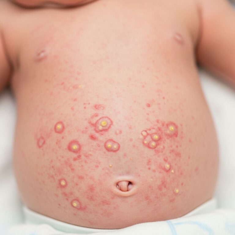
- Non-inflammatory: Asymptomatic scaling mimicking seborrheic dermatitis; loose white ‘cigarette ash’ scales; black dot hairs (T. tonsurans); diffuse ‘carrier state’ dandruff.
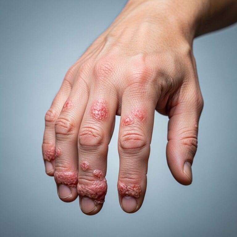
- Inflammatory: Boggy, pus-filled kerion (hypersensitivity reaction); painful swelling, crusting, lymphadenopathy; secondary bacterial infection possible; scarring alopecia if severe.
- Favus: Rare; yellow cups (‘scutula’) on scalp/hairs; T. schoenleinii; malodorous; permanent scarring.
Hair loss manifests as round/oval patches or moth-eaten pattern. Itch mild to severe; pain with kerions. ‘Id reaction’—distant eczematous rash—may occur during treatment. Complications: permanent alopecia (5–10% kerions), abscesses, pyoderma.
| Type | Features | Common Pathogen |
|---|---|---|
| Non-inflammatory | Scaling, black dots, minimal alopecia | T. tonsurans |
| Kerion | Pus-filled swelling, pain, nodes | Zoophilic spp. |
| Favus | Scutula, odor, scarring | T. schoenleinii |
Diagnosis of tinea capitis
Clinical suspicion guides diagnosis, confirmed by:
- Wood’s lamp: Ectothrix green; endothrix dull/no fluorescence; negative in 90% US cases (T. tonsurans).
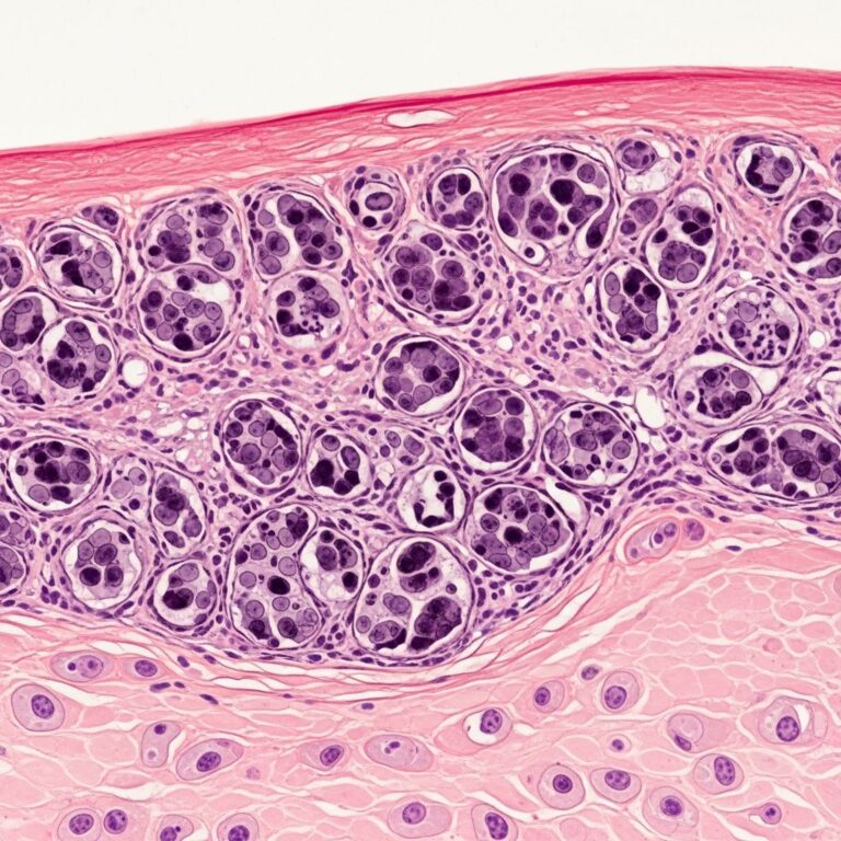
- Hair microscopy: KOH prep shows spores/hyphae; black dots = fractured endothrix hairs.
- Culture: Gold standard; Sabouraud agar; 2–3 weeks; identifies species for treatment tailoring.
- Biopsy: Rarely for kerions/diffuse cases; shows neutrophils, spores in hair.
Do not biopsy routine cases; avoid scalp scraping alone as it misses follicular fungi. Differentiate from alopecia areata, psoriasis, impetigo.
Treatment of tinea capitis
Systemic antifungals essential; topicals inadequate as they don’t penetrate follicles. Duration 4–8 weeks; compliance key to cure (70–90%).
| Drug | Dose (Child) | Duration | Notes |
|---|---|---|---|
| Terbinafine | <20kg: 62.5mg; 20–40kg: 125mg; >40kg: 250mg daily | 4 weeks (T richophyton); 6–8 (Microsporum) | First-line; allylamine; high efficacy, short course. |
| Griseofulvin | 20–25mg/kg/day (microsize/ultramicronized) | 6–8 weeks | Historical first-line; less effective for M. canis; micronized better absorbed. |
| Fluconazole | 6mg/kg daily or 6mg/kg weekly | 3–6 weeks | Alternative; good for resistant cases. |
| Itraconazole | 5mg/kg daily | 2–4 weeks | Pulse options; monitor LFTs. |
Adjuncts: Selenium sulfide/ketoconazole shampoo 2x/week x2 weeks reduces spores/transmission. Kerions: short steroids (prednisone 1mg/kg x2 weeks) + antifungal to prevent scarring.
Monitor LFTs in long courses; cure confirmed by clinical resolution + negative culture (6 weeks post-treatment). Non-responders: culture, check compliance, consider resistance.
What is the outcome for tinea capitis?
Excellent prognosis with early treatment: 80–95% cure. Delays risk kerion (10–20%), scarring alopecia (rare with steroids), chronic carriage. Spores shed months untreated, fueling outbreaks. Relapse from poor compliance/non-simultaneous household Rx. Full hair regrowth 3–6 months; scarring permanent in severe cases.
How can tinea capitis be prevented?
- No sharing combs, hats, towels; clean fomites.
- Screen/treat household/classmates.
- Pet checks; no contact with strays.
- Shampoo all household 2x/week x4 weeks (selenium sulfide).
- Education in schools/endemic areas.
Spore-killing shampoos for carriers; avoid shaving heads (irritates, spreads).
Frequently asked questions (FAQs) on tinea capitis
Q: Is tinea capitis contagious?
A: Yes, highly contagious via direct contact, fomites, animals. Exclude from school/sports until 2–3 weeks treatment/non-infectious.
Q: Can adults get tinea capitis?
A: Rare post-puberty due to sebum, but possible in immunocompromised or close child contact.
Q: Does hair grow back after tinea capitis?
A: Yes, usually fully in 3–12 months unless scarring from kerion.
Q: Can topicals alone treat it?
A: No; systemic required. Shampoos adjunct only.
Q: How long until non-contagious?
A: 1–3 weeks systemic therapy; confirm with culture.
References
- Tinea Capitis: Causes, Symptoms & Treatment — Bosley. 2023. https://www.bosley.com/blog/tinea-capitis-causes-symptoms-treatment/
- Tinea Capitis – StatPearls — NCBI Bookshelf. 2023-10-05. https://www.ncbi.nlm.nih.gov/books/NBK536909/
- Diagnosis and Management of Tinea Infections — American Academy of Family Physicians (AAFP). 2014-11-15. https://www.aafp.org/pubs/afp/issues/2014/1115/p702.html
- Tinea Capitis — DermNet NZ. 2023. https://dermnetnz.org/topics/tinea-capitis
Similar Articles


Read full bio of medha deb