Tinea Capitis: Causes, Symptoms, and Treatment
Understanding scalp ringworm: symptoms, diagnosis, and effective treatment options.

What Is Tinea Capitis?
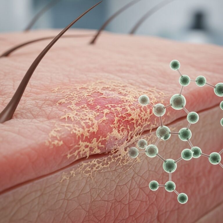
Tinea capitis, commonly known as scalp ringworm or tinea of the scalp, is a fungal infection of the scalp caused by dermatophyte fungi. This contagious condition primarily affects children, though it can occur in adults of any age. The infection develops when fungal organisms colonize the hair shaft and surrounding scalp tissue, leading to inflammation, hair loss, and characteristic symptoms that can be distressing for patients and caregivers.
Unlike other types of ringworm that affect the skin’s surface, tinea capitis penetrates the hair shaft itself, making it more challenging to treat with topical solutions alone. The condition is highly contagious and can spread rapidly in communal settings such as schools and daycare centers, making early recognition and treatment essential.
Understanding the Causative Organisms
Several dermatophyte species can cause tinea capitis, with the specific organism varying geographically. In the United States, Trichophyton tonsurans is responsible for up to 95% of tinea capitis cases, making it by far the most common causative agent in this region. This shift represents a significant change from earlier decades when Microsporum canis was more prevalent.
Geographic variation in causative organisms includes:
- Trichophyton tonsurans — Predominant in the United States (up to 95% of cases)
- Trichophyton violaceum — Dominant organism in Eastern Europe and South Asia
- Microsporum canis — Causes the majority of cases in Africa, Western Europe, Australia, and South America
Risk Factors and Epidemiology
Several factors increase susceptibility to tinea capitis. Age is a significant risk factor, with children experiencing substantially higher infection rates than adults. Children attending daycare centers or schools face increased exposure through close contact with infected peers.
Additional risk factors include:
- Contact with infected pets, particularly those with untreated skin conditions
- Poor personal hygiene practices
- Sharing combs, brushes, hats, or other personal grooming items
- Underlying medical conditions such as diabetes
- Immunodeficiency disorders, including HIV infection
- Prolonged exposure to warm, humid environments that promote fungal growth
The infection spreads readily through direct contact with infected individuals or animals, contaminated surfaces, and shared personal items. Fungal spores can survive on surfaces for extended periods, creating a persistent source of infection.
Clinical Presentation and Symptoms
Tinea capitis presents with variable clinical manifestations depending on the causative organism and host immune response. The most common symptoms include scalp itching, which may range from mild to severe and can significantly impact sleep quality and comfort.
Key clinical features include:
- Bald patches — Circular or irregular areas of hair loss on the scalp
- Scalp inflammation — Redness and irritation of affected areas
- Black dot appearance — Hair loss at the scalp level creating a distinctive stippled pattern, often associated with endothrix infections
- Broken hair — Hair broken 2 to 3 millimeters or more above the scalp surface
- Swollen lymph nodes — Lymphadenopathy in the head and neck region, particularly the occipital and posterior cervical nodes
- Pustules or sores — Secondary bacterial infection or severe inflammation may cause pustule formation
- Kerion formation — A severe inflammatory response characterized by a boggy, pustular mass on the scalp
If left untreated, tinea capitis can result in permanent scarring alopecia and lasting hair loss. The severity and progression depend on the fungal species, immune status of the patient, and duration of infection before treatment initiation.
Diagnosis and Testing
Accurate diagnosis of tinea capitis is essential for appropriate treatment selection. Healthcare providers begin with a comprehensive history and physical examination of the scalp. A dermatologist or primary care physician will evaluate the pattern of hair loss, the appearance of affected areas, and associated symptoms.
Diagnostic methods include:
- Clinical inspection — Visual examination of the scalp and hair for characteristic patterns and lesions
- Wood’s lamp examination — Uses ultraviolet light to identify fluorescent patterns; most Trichophyton species do not fluoresce, but certain Microsporum species show blue-green fluorescence
- Fungal culture — Hair plucking (not cutting) provides the most reliable specimen for culture, allowing organism identification and antifungal susceptibility testing
- Microscopic examination — KOH preparation of hair and scale specimens can demonstrate fungal elements
- Dermoscopy — Advanced visualization technique that can enhance identification of fungal characteristics
When diagnosis is uncertain or for treatment planning purposes, the clinician may obtain a sample by plucking 10 to 15 hairs with follicles or scraping the scalp. Fungal culture remains the gold standard for definitive diagnosis and organism identification.
Treatment Options
Effective treatment of tinea capitis requires systemic antifungal therapy, as topical agents cannot penetrate the hair shaft sufficiently to eradicate infection. The selection of the appropriate antifungal agent depends on the causative organism, patient age, tolerability, and geographic considerations.
Terbinafine
Terbinafine has emerged as a highly effective first-line treatment for tinea capitis in most clinical settings, particularly for Trichophyton tonsurans infections. In 2007, the FDA approved terbinafine oral granules specifically for treating tinea capitis in patients older than four years. This medication demonstrates superior efficacy compared to older agents and offers a shorter treatment duration.
Terbinafine achieves exceptional concentrations in the stratum corneum, reaching levels 75 times higher than plasma concentration after 12 days of therapy. Its long terminal half-life of 200 to 400 hours allows for convenient once-daily dosing and sustained antifungal activity for approximately two months after depletion from plasma. Most clinical trials demonstrate that a four-week course of once-daily terbinafine is effective in treating tinea capitis, with some studies showing efficacy with a two-week course for Trichophyton species infections.
Approved dosing for terbinafine granules is weight-based:
- 125 mg daily for patients weighing less than 25 kg
- 187.5 mg daily for patients weighing 25 to 35 kg
- 250 mg daily for patients weighing 35 kg or more
The treatment duration is typically four to six weeks. Terbinafine offers several advantages over historical treatments, including demonstrated superiority to griseofulvin in terms of efficacy, cost, and shorter treatment duration, with comparable adverse event rates.
Itraconazole
Itraconazole is an effective alternative antifungal agent, particularly for treating infections caused by Microsporum canis. Studies with a combined total of 270 patients demonstrated a 100% cure rate by week 12 using itraconazole at a dose of 5 mg/kg/day. Most patients achieved cure within four to eight weeks of therapy, with equal efficacy reported for both capsule and oral liquid formulations.
Itraconazole is available in multiple formulations and can be administered as continuous daily therapy or pulse therapy, providing treatment flexibility based on individual patient needs and tolerability.
Griseofulvin
Griseofulvin, historically considered the gold standard treatment, has experienced declining use due to documented treatment failures and inferior outcomes compared to newer agents. A retrospective review of medical records revealed a failure rate of 39.3% with griseofulvin treatment. The medication requires an extended treatment duration of eight to twelve weeks, making compliance more challenging, particularly in pediatric patients.
While still available and occasionally used, griseofulvin is no longer recommended as first-line therapy due to superior alternatives with better safety profiles and efficacy rates.
Adjunctive Therapies
Medicated shampoos, while not effective as monotherapy, serve an important role in reducing transmission of infection to other individuals. Antifungal shampoos containing ingredients such as selenium sulfide, zinc pyrithione, or ketoconazole should be used twice weekly during treatment to minimize contagion risk in household and school settings.
Topical antifungal creams have limited effectiveness when used alone but may provide adjunctive benefit for areas with secondary inflammation. Corticosteroid creams should be avoided, as they may worsen the infection by suppressing local immune responses.
Treatment Considerations by Organism Type
| Causative Organism | Geographic Distribution | Recommended First-Line Treatment | Treatment Duration |
|---|---|---|---|
| Trichophyton tonsurans | United States (95% of cases) | Terbinafine | 4 weeks |
| Trichophyton violaceum | Eastern Europe, South Asia | Terbinafine or Itraconazole | 4-6 weeks |
| Microsporum canis | Africa, Western Europe, Australia, South America | Itraconazole | 4-8 weeks |
Prevention Strategies
Preventing tinea capitis transmission requires a multifaceted approach addressing personal hygiene, environmental disinfection, and infection control in communal settings.
Prevention measures include:
- Regular shampooing with thorough scalp cleansing
- Avoiding sharing personal grooming items such as combs, brushes, hairbrushes, and hats
- Using individual towels rather than shared bathroom linens
- Washing towels, bedding, and clothing of infected individuals separately in hot water
- Disinfecting all potentially contaminated surfaces and shared objects
- Seeking veterinary treatment promptly for pets with visible skin infections
- Educating children and caregivers about infection transmission routes
- Maintaining optimal immune function through adequate sleep, nutrition, and stress management
Prognosis and Long-Term Outcomes
With appropriate antifungal treatment, most cases of tinea capitis resolve successfully within the designated treatment period. However, the condition can be challenging to treat, and recurrence occurs in some patients. Compliance with the full treatment course is essential to prevent incomplete cure and subsequent relapse.
Notably, tinea capitis sometimes resolves spontaneously during puberty in adolescents, suggesting that hormonal or immune changes associated with maturation may influence disease progression. However, treatment should not be delayed based on this possibility, as prolonged infection increases the risk of permanent scarring alopecia.
Factors affecting prognosis include the causative organism, immune status of the patient, treatment adherence, and presence of complications such as secondary bacterial infection or severe inflammation.
Frequently Asked Questions
Q: Is tinea capitis contagious?
A: Yes, tinea capitis is highly contagious and spreads readily through direct contact with infected individuals or animals, as well as through shared personal items and contaminated surfaces. Fungal spores can survive on surfaces for extended periods.
Q: Can tinea capitis be treated with only shampoos?
A: No. Medicated shampoos alone are not effective for treating tinea capitis because they cannot penetrate the hair shaft where the fungus resides. Prescription oral antifungal medications are necessary for effective treatment, though antifungal shampoos can help reduce transmission to others.
Q: How long does treatment typically take?
A: Treatment duration varies by medication and organism type. Terbinafine typically requires four weeks of daily dosing, while itraconazole may require four to eight weeks. Older treatments like griseofulvin required eight to twelve weeks of therapy.
Q: Will tinea capitis cause permanent hair loss?
A: If left untreated for prolonged periods, tinea capitis can result in scarring alopecia and permanent hair loss. Prompt diagnosis and appropriate treatment minimize this risk significantly.
Q: Can adults get tinea capitis?
A: While tinea capitis occurs most frequently in children, adults can develop this infection. Risk factors include immunosuppression, exposure to infected individuals or animals, and poor scalp hygiene.
References
- Treatment of Tinea Capitis — PubMed Central, National Institutes of Health. 2019. https://pmc.ncbi.nlm.nih.gov/articles/PMC6615323/
- Tinea capitis — EBSCO Research Starters Health and Medicine. 2024. https://www.ebsco.com/research-starters/health-and-medicine/tinea-capitis
- Ringworm (Tinea Corporis): What It Looks Like, Causes & Treatment — Cleveland Clinic. 2022. https://my.clevelandclinic.org/health/diseases/4560-ringworm
- Dry Scalp: Causes, Treatment & Prevention — Cleveland Clinic. 2023. https://my.clevelandclinic.org/health/symptoms/23326-dry-scalp
- Dermatology for the pediatrician: Advances in diagnosis and management of common skin conditions — Cleveland Clinic Journal of Medicine. 2015. https://www.ccjm.org/content/82/11_suppl_1/S19
Similar Articles


Read full bio of medha deb