Tinea Cruris: Diagnosis, Treatment, And Prevention Guide
Comprehensive guide to jock itch: causes, symptoms, diagnosis, treatment, and prevention strategies for this common fungal groin infection.

What is tinea cruris?
Tinea cruris, commonly known as
jock itch
, is a superficial fungal infection of the groin and adjacent skin areas caused by dermatophytes. These fungi thrive in warm, moist environments, making the intertriginous regions of the groin particularly susceptible. The condition predominantly affects adult males but can occur in females and children as well. It presents as an erythematous, pruritic rash with a characteristic annular pattern, often extending from the inguinal crease onto the upper thighs.The infection is highly contagious and can spread from person to person via direct contact or shared items like towels. It frequently coexists with tinea pedis (athlete’s foot) or onychomycosis (nail fungus), which serve as reservoirs for reinfection. Without proper management, tinea cruris can become chronic, leading to complications such as secondary bacterial infections or lichenification from scratching.
Who gets tinea cruris?
Tinea cruris primarily affects
men
due to the anatomical apposition of the scrotum and thighs, which creates a moist, frictional environment. It is more prevalent in warm climates and among individuals engaging in sports or activities causing excessive sweating. Key risk factors include:- Obesity, leading to skin folds and constant moisture.
- Excessive perspiration (hyperhidrosis).
- Wearing tight, occlusive clothing like synthetic underwear or athletic gear.
- Immunocompromise, diabetes mellitus, or renal/hepatic disease.
- Poor hygiene or sharing contaminated items (towels, locker room benches).
- Concurrent tinea pedis or onychomycosis, acting as fungal reservoirs.
Although less common in women, it can occur in the genital and perianal areas. Children are rarely affected unless overweight or in humid environments. Recurrence is common in summer months due to heat and humidity.
What causes tinea cruris?
Tinea cruris is caused by
dermatophytes
, keratinophilic fungi that infect the stratum corneum. The most common pathogens are:- Trichophyton rubrum (most frequent worldwide).
- Trichophyton mentagrophytes (increasing prevalence in some regions).
- Epidermophyton floccosum and other Trichophyton species.
These fungi spread via direct skin-to-skin contact, fomites, or autoinoculation from feet or nails. The groin area’s warmth (average 32–35°C), moisture from sweat, and friction disrupt the skin barrier, favoring fungal proliferation. Alkaline pH in intertriginous areas further promotes growth. Unlike candidiasis, dermatophytes do not typically invade mucous membranes.
What are the clinical features of tinea cruris?
The hallmark of tinea cruris is a
pruritic, annular plaque
with a raised, scaly border and central clearing. Key clinical features include:- Reddish-brown, sharply demarcated patches starting in the crural folds.
- Extension onto upper inner thighs, buttocks, or perineum (rarely penis or scrotum).
- Intense itching, burning, or stinging.
- Active scaling edge with papules, pustules, or vesicles.
- Bilateral involvement in advanced cases.
Complications may include maceration, secondary candidal or bacterial superinfection, scratch dermatitis, or lichenification. In severe cases, satellite lesions or diffuse erythema occur. Symptoms worsen with heat, exercise, or tight clothing.
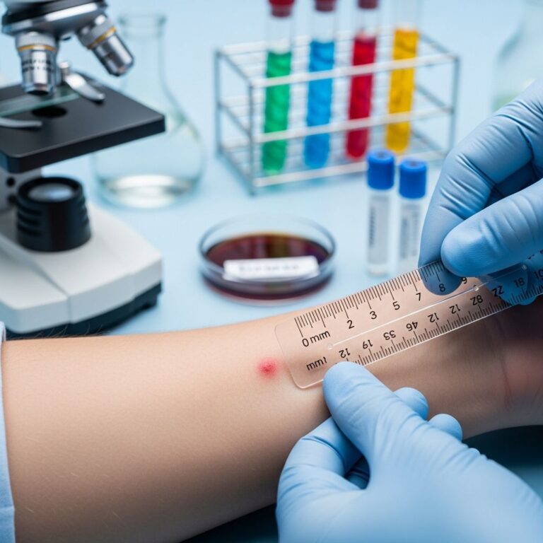
Diagnosis of tinea cruris
Diagnosis is primarily clinical, based on the characteristic rash morphology. Confirmation involves:
- KOH wet mount: Scrapings from the active border show branching hyphae under microscopy.
- Wood’s lamp: Negative (no fluorescence, unlike erythrasma).
- Culture: Sabouraud agar for species identification in atypical or recalcitrant cases.
- Biopsy: Rarely needed, shows fungal elements in stratum corneum.
Differential diagnosis includes candidiasis (satellite pustules, no central clearing), erythrasma (coral-red fluorescence), psoriasis (silvery scales elsewhere), seborrheic dermatitis (greasy scales), or contact dermatitis.
Treatment of tinea cruris
Treatment focuses on topical antifungals, with systemic agents for extensive or resistant cases. Key principles:
- Keep area clean and dry.
- Avoid occlusive clothing; use loose cotton underwear.
- Treat concurrent tinea pedis/onychomycosis to prevent recurrence.
Topical antifungals (first-line)
Apply once or twice daily for 2–4 weeks, extending 2 cm beyond the lesion. Effective agents include:
| Agent | Class | Regimen |
|---|---|---|
| Terbinafine | Allylamine | 1% cream BID x 1–2 weeks |
| Clotrimazole | Azole | 1% cream BID x 2–4 weeks |
| Miconazole | Azole | 2% cream BID x 2 weeks |
| Ketoconazole | Azole | 2% cream QD x 2–4 weeks |
| Econazole | Azole | 1% cream QD x 2 weeks |
Allylamines like terbinafine are fungicidal and often faster-acting. Combination steroid-antifungal creams (e.g., clotrimazole-betamethasone) provide symptomatic relief but risk tinea incognito if overused.
Oral antifungals
Reserved for widespread, chronic, or immunocompromised patients:
- Terbinafine 250 mg daily x 2 weeks.
- Fluconazole 150–200 mg weekly x 2–4 weeks.
- Itraconazole 200 mg daily x 1 week.
Monitor LFTs in hepatic risk patients.
Prevention of tinea cruris
Prevent recurrence with:
- Hygiene: Shower daily, dry thoroughly (use hairdryer on cool), especially after sweating.
- Clothing: Wear breathable cotton underwear; change promptly after exercise.
- Foot care: Treat athlete’s foot; dry feet before groin.
- Avoid sharing: Towels, clothing, or gym equipment.
- Weight management: Reduce skin folds in obesity.
- Prophylactic powders (e.g., miconazole) in high-risk individuals.
Recurrence rates drop significantly with adherence.
Further reading and references
For more on dermatophyte infections, see related topics on tinea pedis and onychomycosis.
Frequently asked questions
Is tinea cruris contagious?
Yes, it spreads via direct contact or fomites. Avoid sharing personal items.
Can women get jock itch?
Yes, though less common, affecting groin and perianal areas.
How long does treatment take?
Topical therapy: 2–4 weeks. Continue 1 week post-clearance.
Does jock itch go away on its own?
Rarely; it often persists or recurs without treatment.
Can I use steroid cream alone?
No, it worsens infection (tinea incognito). Always pair with antifungal.
References
- Tinea Cruris (Jock Itch) — Merck Manuals Professional Edition. 2024. https://www.merckmanuals.com/professional/dermatologic-disorders/fungal-skin-infections/tinea-cruris-jock-itch
- Tinea Cruris — StatPearls, NCBI Bookshelf. 2023-10-16. https://www.ncbi.nlm.nih.gov/books/NBK554602/
- Understanding Tinea Cruris — UMass Memorial Health. 2024. https://www.ummhealth.org/health-library/understanding-tinea-cruris
- Jock Itch (Tinea Cruris) — Cleveland Clinic. 2023-08-01. https://my.clevelandclinic.org/health/diseases/22141-jock-itch-tinea-cruris
- Jock Itch – Symptoms and Causes — Mayo Clinic. 2023-11-13. https://www.mayoclinic.org/diseases-conditions/jock-itch/symptoms-causes/syc-20353807
Similar Articles


Read full bio of Sneha Tete