Tinea Nigra: Causes, Diagnosis, And Treatment Guide
Rare superficial fungal infection causing asymptomatic brown-black patches on palms and soles, often mistaken for melanoma.

Tinea nigra is a rare superficial fungal infection characterised by unilateral, asymptomatic brown or black macules, most commonly on the palms or soles. It is caused by the dematiaceous (pigmented) fungus Hortaea werneckii, previously known as Cladosporium werneckii or Exophiala werneckii, and thrives in tropical and subtropical environments.
What is tinea nigra?
**Tinea nigra** is a superficial phaeohyphomycosis, a type of infection caused by dematiaceous fungi that remain confined to the outermost layer of the skin, the stratum corneum. Unlike true tinea (dermatophyte) infections, it does not invade living tissue or cause inflammation, resulting in painless, non-pruritic patches that can mimic more serious conditions like melanoma.
The infection is extremely superficial and can often be scraped off the skin surface. It predominantly affects individuals in tropical regions but has been reported worldwide, particularly among travelers. The fungus produces a melanin-like pigment, giving the lesions their characteristic dark colour.
Who gets tinea nigra?
Tinea nigra most commonly affects children and young adults, with a slight female predominance in some reports. It is prevalent in tropical and subtropical areas such as Central and South America, Africa, Asia, and coastal Australia. Risk factors include:
- Residence or recent travel to endemic regions
- Hyperhidrosis (excessive sweating), especially on palms and soles
- Contact with soil, sand, sewage, rotting vegetation, wood, or high-salt environments like beaches
- Minor skin trauma, such as cuts or abrasions, facilitating fungal entry
Individuals engaging in gardening, woodworking, or beach activities without protective gloves are at higher risk. The infection is uncommon in temperate climates but can occur via imported cases.
Causes
Tinea nigra is caused by
Hortaea werneckii
, a halophilic (salt-tolerant) black yeast that flourishes in environments with high salinity, acidity, and organic matter, such as sandy beaches, soil, compost, sewage, and decaying wood. Rarely, Stenella araguata has been implicated.Infection occurs through direct inoculation of fungal spores into the stratum corneum via minor trauma. The fungus proliferates in this dead keratin layer without eliciting an immune response, producing darkly pigmented hyphae and yeast cells. Lesions typically appear 2–7 weeks after exposure and remain localised.
Clinical features
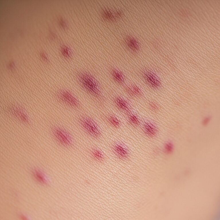
The hallmark of tinea nigra is a
singular or multiple, unilateral, well-demarcated brown to black macule
on the palm (most common), sole, or occasionally fingers, neck, or trunk. Key characteristics include:- Size: 1–5 cm (up to 8 cm in some cases), slowly enlarging
- Shape: Oval, irregular, linear, or serpiginous
- Surface: Slightly scaly or velvety; non-palpable, non-tender
- Symptoms: Asymptomatic; no itch, pain, or inflammation
- Colour: Light brown to jet black, uniform or variegated; persists after washing
Lesions may darken over time due to melanin production. In light-skinned individuals, they appear more striking against pale skin.
Diagnosis
Diagnosis relies on clinical suspicion, history of exposure, and confirmatory tests:
- Clinical examination: A dark macule on acral sites in a tropical traveler raises suspicion.
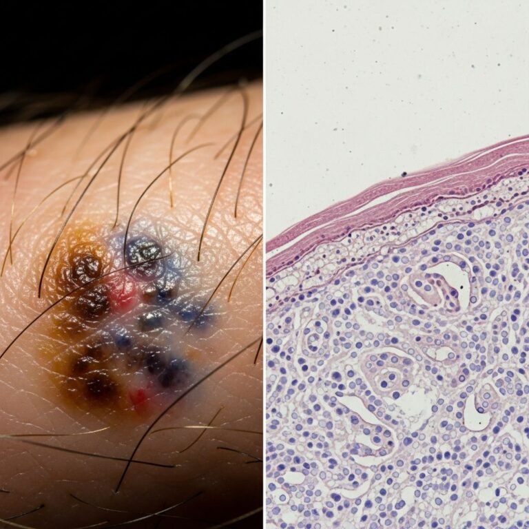
- Dermoscopy: Speckled or dotted pattern with brown-black globules, spoke-wheel-like structures, and disrupted pigment network.
- Microscopy: Skin scrapings in 10–30% KOH show pigmented septate hyphae and round yeast-like cells (‘spaghetti and meatballs’ appearance).
- Culture: Slow-growing (2–3 weeks) on Sabouraud agar; colonies olive-black, velvety.
- Biopsy: Rarely needed; shows hyperkeratosis with pigmented filaments in stratum corneum.
A diagnostic algorithm starts with history and exam, proceeds to KOH prep, and confirms with culture if needed.
Differential diagnosis
Tinea nigra is often misdiagnosed as melanoma or other pigmented lesions. Key differentials include:
| Condition | Distinguishing Features |
|---|---|
| Melanoma | Irregular borders, colour variation, growth; palpable; acral lentiginous type on palms/soles. |
| Lentigo (sun damage, ink spot) | Multiple, symmetrical; history of sun exposure; no scale. |
| Junctional naevus | Uniform colour, stable; uniform globules on dermoscopy. |
| Addison disease | Generalised hyperpigmentation, systemic symptoms. |
| Syphilis (secondary) | Macular rash elsewhere, systemic signs. |
| Pinta/Yaws | Endemic treponemal diseases; hypopigmented centres. |
| Chemical stain (henna, silver nitrate) | History of exposure; fades over time. |
| Post-inflammatory hyperpigmentation | Preceding inflammation or injury. |
Biopsy or excision may be performed if malignancy is suspected.
Treatment
Tinea nigra responds well to topical therapies, with resolution in 2–4 weeks. Options include:
- First-line: Whitfield ointment (3% salicylic acid + 6% benzoic acid) or salicylic acid/urea keratolytics, applied twice daily.
- Antifungals: Topical azoles (ketoconazole 2% cream, miconazole, clotrimazole) or ciclopirox olamine, BID for 2–4 weeks.
- Alternatives: Selenium sulfide shampoo (as lotion), oral itraconazole (200 mg/day for 1–2 weeks) for extensive/large lesions.
- Physical: Curettage or tape stripping to remove infected keratin.
Follow-up confirms clearance; post-inflammatory hyperpigmentation may persist briefly but resolves with keratolytics. Recurrence is rare post-treatment.
What is the outcome of tinea nigra?
Tinea nigra is benign, self-limited if untreated (though persistent), and cures completely without scarring or deeper invasion. Early treatment prevents enlargement and unnecessary biopsies. Hyperpigmentation fades over months. No systemic spread or contagion risk.
Prevention
Preventive measures focus on avoiding inoculation:
- Wear gloves for gardening, woodworking, or soil contact in tropics.
- Avoid barefoot walking on contaminated beaches/sand.
- Maintain foot/palm hygiene, especially if hyperhidrotic.
- Use antifungal powder in at-risk individuals post-exposure.
Awareness reduces misdiagnosis.
Frequently Asked Questions
Q: Is tinea nigra contagious?
A: No, it is not person-to-person transmissible; acquired only from environmental fomites.
Q: Can tinea nigra turn into cancer?
A: No, it is a benign fungal infection; however, its melanoma-like appearance warrants exclusion of malignancy.
Q: How long does treatment take?
A: Typically 2–4 weeks with topical agents; full pigment clearance may take longer.
Q: Does it itch or hurt?
A: No, lesions are entirely asymptomatic.
Q: Can it occur on other body parts?
A: Rarely, yes—neck, trunk, or fingers, but palms/soles predominate.
References
- Tinea nigra — Wikipedia. 2023-10-15. https://en.wikipedia.org/wiki/Tinea_nigra
- Tinea Nigra: Clinical and Diagnostic Guidance — PMC – NIH. 2024-09-01. https://pmc.ncbi.nlm.nih.gov/articles/PMC11380472/
- Tinea nigra — DermNet NZ. 2024-01-01. https://dermnetnz.org/topics/tinea-nigra
- Tinea nigra: Diagnosis, treatment, and remedies — Medical News Today. 2023-05-20. https://www.medicalnewstoday.com/articles/tinea-nigra
- Tinea Nigra: Symptoms, Causes, Treatment, and More — WebMD. 2024-03-10. https://www.webmd.com/skin-problems-and-treatments/what-is-tinea-nigra
Similar Articles


Read full bio of medha deb