Tinea Nigra: 4 Diagnostic Steps To Confirm Diagnosis
Superficial fungal infection causing dark patches on palms and soles, common in tropical climates.

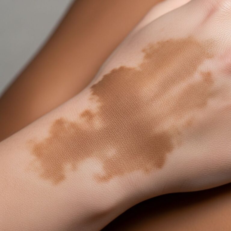
Tinea nigra is a superficial fungal infection characterised by unilateral, asymptomatic, slowly expanding brown to black macules, usually on the palm or fingers but sometimes on the neck, trunk or face.
What is tinea nigra?
**Tinea nigra** is a superficial phaeohyphomycosis caused by the dematiaceous fungus *Hortaea werneckii* (formerly *Exophiala werneckii*, *Cladosporium werneckii* or *Phaeoannellomyces werneckii*). It is a rare condition, also known as pityriasis nigra, keratomycosis nigricans or microsporiosis nigricans. The fungus resides in the soil and is most prevalent in tropical and subtropical climates.
The infection is acquired when fungal spores from soil, decaying vegetation, wood, or compost enter small cuts or abrasions on the skin, particularly in individuals with hyperhidrosis (excessive sweating). It thrives in warm, humid environments and is not contagious from person to person.
Who gets tinea nigra?
Tinea nigra affects all ages and skin types but is more common in:
- Children and young adults
- Individuals living in or travelling to tropical/subtropical regions (e.g., Central/South America, Africa, Asia, Southeast US)
- People with hyperhidrosis
- Those engaged in activities exposing skin to soil/wood, like gardening without gloves
Recent systematic reviews report over 200 cases globally, with a female predominance in some series, though data is limited by underreporting in endemic areas.
What causes tinea nigra?
*Hortaea werneckii* is the primary causative agent, a halophilic (salt-tolerant) dematiaceous fungus producing melanin, giving lesions their characteristic dark colour. Rarely, other fungi like *Stenella araguaya*, *Cyphellophora ludoviensis* or *Exophiala* species have been implicated.
The fungus colonises the outermost layer of skin (stratum corneum), growing as short, septate, pigmented hyphae and spores visible on microscopy. It does not invade deeper tissues, explaining its superficial, benign nature.
What are the signs and symptoms of tinea nigra?
- Unilateral, well-demarcated, hyperpigmented macule (1–4 cm, up to 6 cm)
- Brown to black colour, non-blanching
- Slightly scaly surface (fine, adherent scales)
- Oval, irregular, linear or geographic shape
- Most common on palmar aspect or fingers; less often soles, neck, trunk, face
- Asymptomatic (no itch, pain or inflammation)
- Slow, centrifugal expansion over months/years
Lesions mimic ink stains and may be overlooked until growing larger.
How is tinea nigra diagnosed?
Diagnosis combines clinical suspicion with confirmatory mycology:
- Clinical examination: Dark macule on palm/sole in at-risk patient
- Dermoscopy: Speckled globules, dotted vessels, pigment network or comedo-like openings
- Microscopy: 10–20% KOH preparation reveals pigmented septate hyphae and round/rectangular spores (‘spaghetti and meatballs’ appearance)
- Culture: Slow-growing (2–3 weeks) on Sabouraud dextrose agar at 25–30°C; black, velvety colonies with yeast-to-mould transition
Histology (if needed) shows hyperkeratosis with fungal elements in stratum corneum (periodic acid-Schiff or Fontana-Masson positive).
What is the differential diagnosis for tinea nigra?
| Condition | Key Differentiating Features |
|---|---|
| Melanoma | History of change, asymmetry, irregular borders/colour, >6 mm, elevation; biopsy needed |
| Junctional melanocytic naevus | Uniform colour, stable size, often multiple; uniform dermoscopy globules |
| Lentigo/Lentigo maligna | Multiple lesions, photodamaged skin; lacks scales |
| Chemical/trauma pigmentation | History of exposure (henna, silver nitrate); resolves with time |
| Addison disease | Generalised hyperpigmentation, systemic symptoms |
| Secondary syphilis/pinta/yaws | Multiple lesions, systemic signs, positive serology |
| Post-inflammatory hyperpigmentation | Preceding inflammation, resolves gradually |
Related Stories


Biopsy is warranted for atypical/diagnostic uncertainty cases to exclude melanoma.
What is the treatment for tinea nigra?
Topical antifungals are first-line, achieving 100% resolution in systematic reviews (average 4 weeks). Options include:
- Whitfield ointment (benzoic/pericylic acid): BID 2–4 weeks (12/100+ cases)
- Ketoconazole 2% cream: BID 2–4 weeks (11 cases)
- Terbinafine 1% cream/Isoconazole 1% cream: BID 2–4 weeks (10 cases each)
- Other azoles (miconazole, clotrimazole, econazole): BID until clear
- Selenium sulfide lotion (2.5%): Daily 2 weeks
- Keratolytics (salicylic acid, urea): Soften hyperkeratosis
Mechanical debridement (curettage) may suffice alone. Oral itraconazole/fluconazole reserved for extensive/recalcitrant cases (rare).
Post-treatment hyperpigmentation fades with time or keratolytics.
What is the outcome for tinea nigra?
Excellent prognosis with treatment; lesions resolve completely without scarring. Recurrence is rare post-eradication. Untreated lesions persist indefinitely but remain superficial/benign.
Spontaneous resolution is exceptional (2 cases reported).
How can tinea nigra be prevented?
- Wear gloves for gardening/soil contact in tropics
- Avoid minor injuries in endemic areas
- Practice good foot/palm hygiene if hyperhidrotic
- Early treatment of suspicious lesions
Frequently Asked Questions (FAQs)
Is tinea nigra contagious?
No, it is not person-to-person transmissible; acquired from environment.
Can tinea nigra occur on other body parts?
Rarely neck, trunk, face or neck; palms/soles predominate.
Does tinea nigra itch or hurt?
No, it is asymptomatic.
Is tinea nigra a type of ringworm?
No, despite ‘tinea’ name; caused by mould, not dermatophyte.
How long does treatment take?
Typically 2–4 weeks with topical antifungals.
Can I treat tinea nigra at home?
Confirm diagnosis first; use OTC antifungals like clotrimazole after doctor advice.
References
- Tinea nigra — DermNet NZ. 2023. https://dermnetnz.org/topics/tinea-nigra
- Systematic Review of Tinea Nigra: A Clinical Approach — PMC/NCBI. 2024-10-15. https://pmc.ncbi.nlm.nih.gov/articles/PMC12028572/
- Tinea nigra — Wikipedia (sourced from primary refs). 2025. https://en.wikipedia.org/wiki/Tinea_nigra
- Tinea nigra: Diagnosis, treatment, and remedies — Medical News Today. 2024. https://www.medicalnewstoday.com/articles/tinea-nigra
- Tinea Nigra: Symptoms, Causes, Treatment — WebMD. 2024. https://www.webmd.com/skin-problems-and-treatments/what-is-tinea-nigra
Similar Articles


Read full bio of Sneha Tete