Toxic Epidermal Necrolysis: Causes, Symptoms, and Treatment
Understanding TEN: A severe skin reaction requiring immediate medical intervention and specialized care.

What Is Toxic Epidermal Necrolysis?
Toxic epidermal necrolysis (TEN) is a rare but potentially life-threatening skin disorder characterized by extensive exfoliation of the epidermis and mucous membranes. This severe cutaneous hypersensitivity reaction causes the skin to blister and peel in sheets, leaving large, raw areas exposed. The condition represents one of the most serious dermatological emergencies, requiring immediate hospitalization and intensive medical care.
TEN is closely related to Stevens-Johnson syndrome (SJS), with the primary distinction being the extent of skin involvement. While both conditions share similar causes, symptoms, and treatment approaches, TEN involves more extensive detachment of the epidermis, typically affecting greater than 30% of the body surface area. The rapid progression and severe complications make early recognition and treatment critical for patient survival.
Understanding the Causes of Toxic Epidermal Necrolysis
Medications are the most common triggers of toxic epidermal necrolysis, accounting for the majority of cases. The condition typically develops within the first 8 weeks of using a new medication, though it can occasionally occur after prolonged use.
Medication-Related Causes
Several classes of medications have been implicated in TEN development:
- Sulfonamide antibiotics and other sulfa drugs (such as sulfasalazine)
- Anticonvulsant medications used to treat seizures
- Aminopenicillin antibiotics, including ampicillin and amoxicillin
- Fluoroquinolone and other broad-spectrum antibiotics
- Cephalosporin antibiotics
- Nonsteroidal anti-inflammatory drugs (NSAIDs)
- Medications for gout treatment
- Antiretroviral medications used in HIV treatment
Non-Medication Causes
While less common, TEN can occasionally result from causes other than medications. These include infections such as Mycoplasma pneumoniae or cytomegalovirus, vaccines, herbal medicine use, and contact with certain chemicals. In some cases, no identifiable trigger is found, suggesting a possible genetic predisposition in susceptible individuals.
The Pathophysiology of Toxic Epidermal Necrolysis
The exact mechanisms underlying toxic epidermal necrolysis involve complex immune system interactions. Research indicates that granulysin, a protein released from cytotoxic T cells and natural killer cells, plays a significant role in keratinocyte death. The concentration of granulysin in blister fluid correlates directly with disease severity. Additionally, interleukin-15, an inflammatory cytokine, has been found to be elevated in patients with TEN and stimulates granulysin production.
Another proposed mechanism involves Fas, a cell-surface receptor that induces programmed cell death (apoptosis). When Fas interacts with its ligand, particularly a soluble form released from mononuclear cells, it triggers cell death and contributes to blister formation. Genetic predisposition has also been suggested as a contributing factor, as certain individuals appear more susceptible to developing TEN when exposed to triggering medications.
Recognizing Symptoms and Clinical Presentation
Toxic epidermal necrolysis typically follows a predictable progression, though symptoms can vary among individuals. Prompt recognition of early signs is essential for initiating timely treatment.
Initial Prodromal Phase
The condition usually begins 1 to 3 weeks after starting the offending medication. Patients first experience flu-like symptoms lasting 1 to 3 days before skin manifestations appear, including:
- Malaise and general feeling of illness
- High fever
- Headache
- Cough
- Keratoconjunctivitis (eye inflammation)
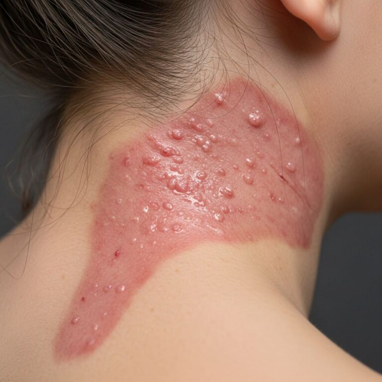
Dermatological Manifestations
Following the prodromal phase, characteristic skin lesions develop suddenly:
- Red macules often appearing in a target configuration
- Rapid spread and coalescence of lesions into large flaccid bullae
- Blistering and peeling of skin in sheets without necessarily requiring prior blistering
- Large, raw areas of exposed skin
- Lesions typically appearing first on the face, neck, and upper trunk before spreading
- Sloughing of skin over 1 to 3 days
Mucosal and Systemic Involvement
Beyond skin manifestations, patients frequently experience painful blistering, redness, and discomfort affecting:
- Eyes and eyelids
- Mouth and throat
- Genitals and urethral area
- Anal area
Additional symptoms may include photophobia (pain with light exposure), fatigue, muscle pain, joint pain, and difficulty swallowing. The rapid disease progression, typically occurring within three days, underscores the urgency of medical intervention.
Diagnosis of Toxic Epidermal Necrolysis
Diagnosis is usually apparent from the clinical presentation and characteristic appearance of initial lesions. The combination of the distinctive target-shaped macules, rapid progression to widespread blistering and skin sloughing, and recent medication exposure typically provides sufficient diagnostic information. A detailed medication history and timeline of symptom onset are critical diagnostic elements.
In some cases, skin biopsy may be performed to confirm the diagnosis and rule out other conditions. Histopathological examination typically reveals full-thickness epidermal necrosis with minimal inflammation, helping distinguish TEN from similar conditions like erythema multiforme.
Treatment and Management Strategies
Successful treatment of toxic epidermal necrolysis requires early recognition and rapid intervention in specialized care settings. The disease progresses rapidly, making immediate hospitalization essential.
Immediate Management Steps
The cornerstone of early management involves two critical actions:
- Immediate discontinuation of the suspected offending medication
- Early referral to a burn unit or intensive care unit with specialized experience in managing severe skin conditions
Research demonstrates that these measures, when implemented within the first 24 hours of blister formation, significantly decrease infection rates, reduce hospital stays, and improve overall survival rates.
Supportive Care Measures
The primary treatment approach focuses on supportive care while the skin undergoes re-epithelialization. Essential supportive measures include:
- Aggressive fluid resuscitation through intravenous access
- Electrolyte and mineral replacement
- Comprehensive pain management
- Meticulous wound care and protective bandaging
- Nutritional support, including nasogastric tube feeding if necessary
- Isolation protocols to prevent secondary infections
- Antibiotic therapy as appropriate
Specialized Medical Interventions
Beyond basic supportive care, several specialized treatments may be employed:
- Intravenous immunoglobulin (IVIG) to prevent further immune system damage
- Intravenous corticosteroids, though their role remains somewhat controversial
- Cyclosporine, an immunosuppressive agent that may limit disease progression
- Plasmapheresis to remove circulating immune factors
- Tumor necrosis factor (TNF)-alpha inhibitors
Specialized Care Requirements
Treatment in a burn unit or intensive care unit setting is typically necessary because the management approach closely parallels treatment for extensive burn injuries. Patients may require eye care with daily examination, eyelid cleaning, and lubrication to prevent corneal damage. Ophthalmologic consultation is recommended for all TEN patients to assess and manage eye involvement.
Prognosis and Long-Term Outcomes
The prognosis for toxic epidermal necrolysis depends on disease severity, extent of skin involvement, patient age, and timing of treatment initiation. Severe TEN resembles extensive burns, with patients experiencing acute illness, potential inability to eat or open their eyes, and massive fluid and electrolyte losses.
Mortality and Survival Rates
Mortality rates vary significantly based on disease severity and treatment timing. In adults, mortality can range from 25 to 35%, though rates tend to be lower in children and when treatment is initiated early. The most common causes of death include infection, multiorgan failure, and sepsis resulting from loss of skin barrier function.
Potential Complications
Survivors of toxic epidermal necrolysis may experience significant long-term complications:
- Permanent skin color changes and dyspigmentation
- Abnormal mole growth (dysplastic nevi)
- Abnormal growth and changes to fingernails and toenails
- Hair loss
- Skin scarring, particularly following severe infection
- Corneal scarring and vision changes
- Oral cavity changes including periodontal disease
- Lung damage and pulmonary complications
- Chronic dry eyes and other ocular complications
Prevention and Risk Mitigation
While toxic epidermal necrolysis cannot always be prevented, certain measures can reduce risk. Patients with a history of TEN or Stevens-Johnson syndrome should maintain a detailed record of all medications that triggered or are suspected of triggering the reaction. Healthcare providers should review this list before prescribing new medications, particularly avoiding structurally similar drugs.
Patients beginning new medications should be educated about warning signs and instructed to seek immediate medical attention if fever, rash, or mucosal involvement develops. Close monitoring during the first 8 weeks of new medication use is particularly important, as this is the highest-risk period for TEN development.
When to Seek Emergency Medical Care
Immediate medical evaluation is warranted if any of the following occur:
- Development of skin problems, rash, or blistering after starting a new medication
- High fever combined with skin changes
- Rapid spread of painful red areas on the skin
- Widespread blistering or skin peeling
- Pain or involvement of eyes, mouth, throat, or genitals
- Flu-like symptoms followed by rash development
Frequently Asked Questions About Toxic Epidermal Necrolysis
Q: How quickly does toxic epidermal necrolysis develop?
A: TEN typically develops within 1 to 3 weeks after starting an offending medication. Initial flu-like symptoms appear first, followed by skin manifestations that progress rapidly over 1 to 3 days.
Q: Is toxic epidermal necrolysis contagious?
A: No, toxic epidermal necrolysis is not contagious. It is a medication reaction or immune response, not an infectious disease. However, patients are at high risk of developing secondary infections due to loss of skin barrier function, so isolation may be recommended to protect patients from external pathogens.
Q: Can toxic epidermal necrolysis recur?
A: While TEN typically occurs once, there is a risk of recurrence if a patient is exposed to the same medication or structurally similar drugs. Patients should maintain accurate records of triggering medications and inform all healthcare providers.
Q: What is the difference between toxic epidermal necrolysis and Stevens-Johnson syndrome?
A: Both conditions are severe cutaneous hypersensitivity reactions with similar causes and treatment. The primary difference is the extent of skin involvement—TEN affects more than 30% of body surface area, while SJS affects less than 10%, with overlapping cases (10-30%) classified as SJS/TEN.
Q: How long does recovery from toxic epidermal necrolysis take?
A: Recovery can take weeks to months depending on disease severity and extent of skin involvement. Complete re-epithelialization of affected areas requires time, and patients may experience ongoing complications during recovery.
Q: Are there any medications that should never be used after TEN?
A: Patients should permanently avoid the specific medication that triggered TEN and any structurally similar drugs. Healthcare providers can help identify safe alternative medications for treating underlying conditions.
References
- Stevens-Johnson Syndrome (SJS) and Toxic Epidermal Necrolysis (TEN) — Merck Manuals Professional Edition. 2024. https://www.merckmanuals.com/professional/dermatologic-disorders/hypersensitivity-and-reactive-skin-disorders/stevens-johnson-syndrome-sjs-and-toxic-epidermal-necrolysis-ten
- Toxic Epidermal Necrolysis — National Center for Biotechnology Information, StatPearls. 2024. https://www.ncbi.nlm.nih.gov/books/NBK574530/
- Toxic Epidermal Necrolysis in Children — University of Rochester Medical Center. 2024. https://www.urmc.rochester.edu/encyclopedia/content?contenttypeid=90&contentid=p01911
- Toxic Epidermal Necrolysis — Texas Children’s Hospital. 2024. https://www.texaschildrens.org/content/conditions/toxic-epidermal-necrolysis
- Stevens-Johnson Syndrome — Mayo Clinic. 2024. https://www.mayoclinic.org/diseases-conditions/stevens-johnson-syndrome/symptoms-causes/syc-20355936
- Stevens-Johnson Syndrome — National Health Service (NHS). 2024. https://www.nhs.uk/conditions/stevens-johnson-syndrome/
- Toxic Epidermal Necrolysis — Boston Children’s Hospital. 2024. https://www.childrenshospital.org/conditions/toxic-epidermal-necrolysis
Similar Articles


Read full bio of medha deb