Toxic Epidermal Necrolysis Pathology: Key Diagnostic Features
Comprehensive pathology of TEN: from clinical features and histopathology to detailed microscopic findings and differential diagnosis.

Author: Dr. Harriet Cheng, Dermatopathologist, Reviewed: Dr. Ian Hay, Dermatologist
Introduction
Toxic epidermal necrolysis (TEN) represents the severe end of the StevensJohnson syndrome (SJS) / TEN spectrum, characterized by extensive epidermal necrosis and detachment affecting more than 30% of the body surface area (BSA). This life-threatening mucocutaneous reaction is most commonly triggered by medications, leading to massive keratinocyte apoptosis through immune-mediated mechanisms. The condition carries a mortality rate of 2550%, primarily due to sepsis, multiorgan failure, and respiratory complications. Understanding the pathology of TEN is crucial for prompt diagnosis, differentiation from similar entities, and guiding intensive supportive care.
This article details the clinical presentation, histopathological features, electron microscopy findings, direct immunofluorescence (DIF) patterns, and differential diagnosis of TEN, providing dermatologists and pathologists with comprehensive diagnostic criteria.
Clinical features
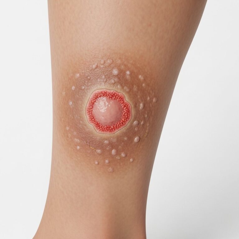
TEN typically begins with a prodromal phase of fever, malaise, sore throat, and cough 13 weeks after exposure to the culprit drug, such as anticonvulsants, sulfonamides, allopurinol, or NSAIDs. This is followed by rapid onset of painful erythematous macules, atypical target lesions, and flaccid blisters progressing to sheet-like epidermal detachment involving >30% BSA.
- Positive Nikolsky sign: Lateral pressure on erythematous skin causes epidermal sloughing due to basal keratinocyte fragility.
- Mucosal involvement: Nearly 100% of cases show oral, ocular, and genital erosions; eye involvement ranges from conjunctivitis to corneal sloughing.
- Cutaneous progression: Lesions start on trunk and face, spreading centrifugally; severe skin pain precedes visible detachment.
- Systemic signs: High fever, tachycardia, hypotension, and risk of sepsis from skin barrier loss.
Severity is classified by BSA detachment: SJS (<10%), SJS/TEN overlap (1030%), TEN (>30%). Early recognition prevents progression and complications like acute respiratory distress syndrome.
Histopathology
Skin biopsy is essential for confirming TEN diagnosis, showing characteristic full-thickness epidermal necrosis with minimal inflammation. The hallmark is widespread keratinocyte apoptosis progressing from scattered “satellite” necrosis to confluent coagulative necrosis of the entire epidermis.
Low power
At scanning magnification, biopsies reveal extensive epidermal detachment, with full-thickness necrosis creating a subepidermal split. Sparse perivascular lymphocytic infiltrate is present in the superficial dermis, without vasculopathic changes. Detached necrotic epidermis appears as pale eosinophilic sheets above a relatively preserved dermis.
High power
- Early changes: Scattered apoptotic keratinocytes (round, eosinophilic bodies with pyknotic nuclei) at basal layer, expanding to suprabasal zones.
- Established TEN: Confluent full-thickness necrosis with ghost-like keratinocytes, loss of nuclear detail, and eosinophilic cytoplasm.
- Dermal infiltrate: Sparse superficial perivascular lymphocytes; rare eosinophils or neutrophils.
- No significant vacuolar change: Distinguishes from interface dermatitis; basal vacuolation is minimal despite extensive necrosis.
Absent or minimal dermal inflammation underscores the primarily cytotoxic epidermal process driven by granulysin, FasL, and perforin/granzyme pathways.
Electron microscopy
Ultrastructural examination reveals early keratinocyte injury before light microscopic necrosis becomes evident. Key findings include:
- Cytoplasmic vacuolization: Basal keratinocytes show perinuclear vacuoles and swollen mitochondria.
- Desmosomal degeneration: Widened intercellular spaces and detached desmosomes lead to intraepidermal cleavage.
- Apoptotic bodies: Condensation of nuclear chromatin and cytoplasmic fragmentation.
- Basal lamina preservation: Split occurs above intact basement membrane, explaining subepidermal blister appearance.
These changes reflect Fas-mediated apoptosis and cytotoxic T-cell/NK cell attack via granulysin release, confirming immune pathogenesis.
Direct immunofluorescence
DIF is negative or non-specific in TEN, helping exclude autoimmune bullous diseases.
| Finding | Description |
|---|---|
| Intercellular IgM | Focal, non-specific; represents necrotic debris |
| Basement membrane zone | Negative for linear IgG/IgA/C3; rules out bullous pemphigoid |
| Cytoplasmic staining |
Negative DIF supports drug-induced cytotoxic mechanism over autoantibody-mediated blistering.
Differential diagnosis
TEN must be distinguished from mimics with epidermal necrosis or detachment. Key differentiators:
| Condition | Key Distinguishing Features |
|---|---|
| Staphylococcal scalded skin syndrome (SSSS) | Intraepidermal (granular layer) split; superficial; children; +ve Tzanck smear for acantholytic cells; recovers quickly |
| Erythema multiforme major | Typical targets; sparse necrosis; prominent vacuolar interface; less detachment |
| Graft-versus-host disease | Apoptosis confined to basal layer; vacuolar degeneration; lymphocytic satellitosis |
| Pemphigus vulgaris | Suprabasal acantholysis; +ve DIF (IgG intercellular) |
| AGEP | Subcorneal pustules; eosinophils; rapid onset post-antibiotics |
| MTX toxicity | Epidermal atrophy; pancytopenia; no T-cell markers; high-dose MTX history |
Pathogenesis
TEN results from CD8+ T-cell and NK-cell mediated cytotoxicity triggered by drugs presenting as haptens. Key mediators:
- Granulysin: Primary effector; 9-kDa protein causing keratinocyte lysis
- Fas-FasL: Death receptor pathway inducing apoptosis
- Perforin/granzyme B: Pore formation and caspase activation
- Genetic risk: HLA associations (e.g., HLA-B*15:02 with carbamazepine)
Management implications
Pathological confirmation guides withdrawal of culprit drug, transfer to burn ICU, fluid resuscitation, infection control, and potential immunosuppressants (IVIG, cyclosporine). Prognosis uses SCORTEN score; mortality correlates with BSA and age.
Frequently asked questions
Q: What is the hallmark histopathology of TEN?
A: Full-thickness epidermal necrosis with sparse dermal lymphocytic infiltrate and subepidermal detachment.
Q: How does TEN differ from SSSS pathologically?
A: TEN shows full-thickness necrosis and subepidermal split; SSSS has superficial (granular layer) cleavage.
Q: Is DIF positive in TEN?
A: No, DIF is negative or shows non-specific IgM; used to exclude pemphigus/bullous diseases.
Q: What mediates keratinocyte death in TEN?
A: Granulysin from cytotoxic T-cells/NK cells, plus FasL and perforin/granzyme pathways.
Q: When is biopsy indicated in suspected TEN?
A: Always for confirmation, especially to differentiate from mimics; use 12 mm punch from perilesional skin.
References
- Toxic Epidermal Necrolysis (TEN): Risk Factors, Clinical Features — The Plastics Fella. 2023. https://www.theplasticsfella.com/toxic-epidermal-necrolysis-ten/
- Toxic Epidermal Necrolysis: A Review of Past and Present — Skin Therapy Letter. 2022-06-15. https://www.skintherapyletter.com/dermatology/toxic-epidermal-necrolysis/
- A review on toxic epidermal necrolysislike superficial wound lesions — PMC (NCBI). 2024. https://pmc.ncbi.nlm.nih.gov/articles/PMC11646660/
- TEN – Symptoms and causes — Mayo Clinic. 2025-01-15. https://www.mayoclinic.org/diseases-conditions/toxic-epidermal-necrolysis/symptoms-causes/syc-20491903
- Toxic Epidermal Necrolysis – StatPearls — NCBI Bookshelf. 2024-10-20. https://www.ncbi.nlm.nih.gov/books/NBK574530/
- Stevens Johnson Syndrome / Toxic Epidermal Necrolysis — DermNet NZ. 2023-05-01. https://dermnetnz.org/topics/stevens-johnson-syndrome-toxic-epidermal-necrolysis
Similar Articles


Read full bio of Sneha Tete