Toxoplasmosis Pathology: What Clinicians Need To Know
Comprehensive pathology of Toxoplasma gondii infection: from histopathology to clinical manifestations and diagnosis.

Toxoplasmosis is a zoonotic infection caused by the obligate intracellular protozoan parasite Toxoplasma gondii, which infects up to one-third of the global human population. While often asymptomatic in immunocompetent individuals, it can lead to severe systemic disease in immunocompromised patients or congenital infections. Pathology primarily involves tissue cysts and tachyzoites, with rare cutaneous manifestations.
Authoritative Facts
- T. gondii life cycle includes oocysts from feline definitive hosts, shed in feces, and tissue cysts in intermediate hosts like humans via undercooked meat or contaminated water.
- Intracellular tachyzoites cause acute cytopathic effects, inflammation, and necrosis; bradyzoites form latent cysts in brain, muscle, and eyes.
- Cell-mediated immunity, particularly IFN-γ, controls infection; defects lead to reactivation.
- Skin involvement is rare, mostly in immunocompromised, presenting as ulcers, maculopapular eruptions, or nodules.
What is Toxoplasmosis?
Toxoplasmosis results from infection with Toxoplasma gondii, a cosmopolitan parasite. In humans, primary infection is usually subclinical, but dissemination occurs via tachyzoites crossing barriers like the blood-brain barrier. Latent cysts persist lifelong in neural and muscular tissues. Reactivation in immunosuppression causes encephalitis, pneumonitis, or retinitis.
Transmission routes include oral ingestion of oocysts from cat feces-contaminated soil/food, tissue cysts in undercooked meat (pork, lamb), or transplacentally. Rare vectors include blood transfusion or organ transplant.
Who Gets Toxoplasmosis?
Global seroprevalence varies: 10-30% in Europe/North America, up to 80% in parts of Africa/Latin America. Risk factors include eating raw/undercooked meat, unwashed vegetables, gardening without gloves, or HIV (CD4 <100 cells/μL).
Immunocompetent hosts rarely develop severe disease; symptoms mimic mononucleosis. Immunocompromised (AIDS, transplant, chemotherapy) face life-threatening dissemination. Pregnant women risk fetal transmission, worse in first trimester.
Histopathology / Pathology
Key pathologic hallmarks are tachyzoites (acute phase: crescent-shaped, 2-6 μm, intracytoplasmic) and bradyzoites (chronic cysts: 10-100 μm, PAS-positive, containing thousands of organisms). Tachyzoites cause direct cell lysis, recruiting macrophages/neutrophils via IL-12/IFN-γ.
Cutaneous pathology (rare): Dense dermal infiltrates of lymphocytes, plasma cells, multinucleated giants, focal abscesses. PAS/Giemsa stains reveal tachyzoites in epidermis/dermis. Seen in isolated ulcers (even immunocompetent) or roseola-like eruptions.
| Feature | Acute (Tachyzoites) | Chronic (Cysts) |
|---|---|---|
| Morphology | Crescentic, banana-shaped | Spherical, thick-walled |
| Size | 2×7 μm | 10-100 μm |
| Stain | Giemsa, Wright (basophilic) | PAS positive |
| Location | Any cell | Neurons, myocytes |
Brain: Perivascular cuffing, microglial nodules, necrosis. Muscle: Cysts in sarcoplasm, minimal inflammation unless ruptured.
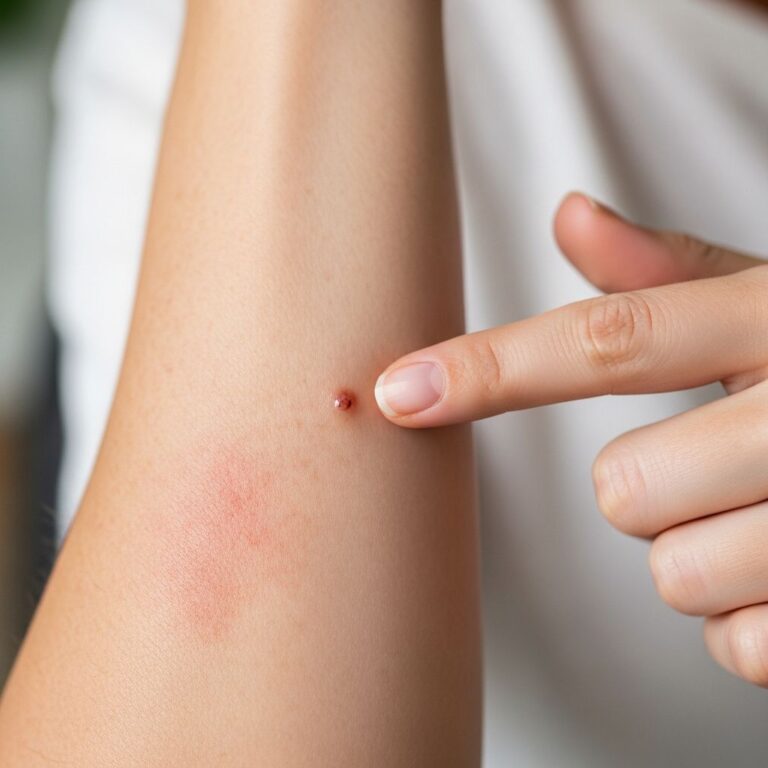
Clinical Features of Cutaneous Toxoplasmosis
Skin Lesions of Congenital Toxoplasmosis
- Maculopapular rash, petechiae, jaundice (hydrops-like).
- Rare strawberry tongue, vesicles.
Skin Lesions of Acute Acquired Toxoplasmosis
- Immunocompetent: Transient maculopapular (trunk/extremities), urticaria, erythema multiforme, erythroderma (rare).
- Immunocompromised: Ulcers/nodules (face, trunk), panniculitis, Kaposi-like.
A case of primary cutaneous toxoplasmosis: 64-year-old female with nonhealing dorsal hand ulcer (1 year), no systemic symptoms/seropositivity. Biopsy showed plasma cells, giants, PAS+ tachyzoites—resolved post-excision.
Diagnosis
Serology: IgM (acute), IgG (past/chronic). Avidity tests distinguish recent vs. old. PCR on blood/amniotic fluid/CSF for active disease. Histopathology confirmatory: tachyzoites/cysts in biopsy.
For congenital: Fetal ultrasound (hydrocephalus, calcifications), amniocentesis PCR. Eye exam for chorioretinitis.
Treatment
Treat active/immunocompromised/congenital cases. First-line: Pyrimethamine + sulfadiazine + folinic acid (tachyzoites only; cysts persist).
- Allergic: Clindamycin or atovaquone substitute.
- Prophylaxis (HIV): TMP-SMX daily.
- Pregnancy: Spiramycin (prevent transmission); post-transmission, pyrimethamine combo from 18 weeks.
Duration: 4-6 weeks acute; lifelong suppression in AIDS.
Frequently Asked Questions (FAQs)
Q: Can toxoplasmosis cause skin ulcers in healthy people?
A: Extremely rare, but documented in immunocompetent adults as nonhealing ulcers with tachyzoites on biopsy.
Q: How is cutaneous toxoplasmosis diagnosed?
A: Biopsy with H&E/PAS/Giemsa showing parasites; serology often negative in isolated cases.
Q: Is toxoplasmosis curable?
A: Acute tachyzoites yes, but latent cysts no—risk of reactivation.
Q: What prevents congenital toxoplasmosis?
A: Pregnant women avoid raw meat, cat litter, wash produce; screen serology early.
Q: Does toxoplasmosis affect the eyes?
A: Yes, chorioretinitis—congenital or reactivated, causing scars/blindness.
Complications
Encephalitis (ring-enhancing lesions), pneumonitis, myocarditis, retinitis. Congenital: Microcephaly, chorioretinitis, seizures.
References
- Toxoplasmosis Presenting as Nonhealing Cutaneous Ulcer — Adhikari et al. 2020-10-01. https://onlinelibrary.wiley.com/doi/10.1155/2020/8874800
- Toxoplasmosis – DermNet — DermNet NZ. 2023. https://dermnetnz.org/topics/toxoplasmosis
- Toxoplasmosis – StatPearls — NCBI Bookshelf. 2023-07-17. https://www.ncbi.nlm.nih.gov/books/NBK563286/
- DPDx – Toxoplasmosis — CDC. 2024. https://www.cdc.gov/dpdx/toxoplasmosis/index.html
- Pathology of toxoplasmosis — YouTube (CDC DPDx). 2018. https://www.youtube.com/watch?v=63tiFHOzYb8
Similar Articles


Read full bio of Sneha Tete