Transient Acantholytic Dermatosis Images
Explore clinical images and detailed insights into Grover disease, its features, diagnosis, and management strategies.

Author: Dr Amanda Oakley, Dermatologist, Hamilton, New Zealand. Reviewed: 2023-10-01
What is transient acantholytic dermatosis?
Transient acantholytic dermatosis is a relatively common acquired skin disease that tends to occur in adult men, particularly over the age of 50 years. Transient acantholytic dermatosis is also known as Grover’s disease. It is characterised by a persistent pruritic (itching) eruption composed of small red spotty papules. The rash is usually located on the trunk (chest, abdomen and back). It can last for months or years, relapsing in the summer months or with excessive sweating.
The cause is unknown, but it is more common in older adults, particularly white men. Risk factors include heat, sweating, sun exposure, bed rest, and certain medications. Histologically, it shows focal acantholysis with dyskeratotic cells, confirming the diagnosis.
Clinical features of transient acantholytic dermatosis
Transient acantholytic dermatosis presents as an acute or chronic rash composed of 1–5 mm firm pink or red-brown papules, which may be flat-topped, crusted or scaly. The rash is usually very itchy and often affects the chest, back and upper abdomen. It may also involve the groin and thighs. The papules may coalesce into larger plaques.
- Sudden onset of pruritic papules on the trunk
- Intensified by heat, sweating, or friction
- May wax and wane seasonally
- Typically spares face, extremities, and mucous membranes
Images of transient acantholytic dermatosis
Figure 1: Grover disease on the chest
Multiple erythematous scaly papules coalescing into plaques on the anterior chest of an older man. The lesions show a crusted surface and are distributed symmetrically across the presternal area.
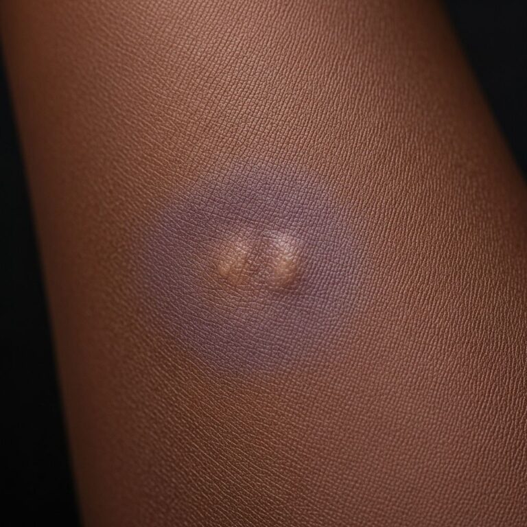
Figure 2: Close-up of Grover disease papules
Close-up view revealing 2-3 mm pink papules with adherent scale and subtle vesiculation. Background erythema highlights the monomorphous nature of the eruption.
Figure 3: Extensive Grover disease on trunk
Widespread involvement of chest, abdomen, and flanks with hundreds of pruritic papules in a 72-year-old hospitalized patient after prolonged bed rest.
Figure 4: Grover disease in skin folds
Lesions concentrated in inframammary folds and abdominal creases, exacerbated by occlusion and perspiration, showing more vesicular morphology.
Figure 5: Partial resolution after treatment
Residual postinflammatory hyperpigmentation following successful cryotherapy, demonstrating fading of active papules with mild PIH macules remaining.
Histopathology of transient acantholytic dermatosis
Skin biopsy is often diagnostic, showing suprabasal acantholysis (loss of cohesion between keratinocytes) with dyskeratotic (corpse-like) cells. Several histological patterns exist: Darier-like, Hailey-Hailey-like, spongiotic, and pemphigus-like. Focal acantholysis remains the hallmark finding.
- Suprabasal clefting with acantholytic cells floating in clefts
- Dyskeratotic keratinocytes with eosinophilic cytoplasm
- Spongiosis in some variants
- No true bullae formation typically
Diagnosis
Diagnosis is primarily clinical but confirmed by histopathology. Key is recognising the characteristic truncal distribution of pruritic papules in at-risk patients (older males, heat exposure). Skin biopsy differentiates from mimics.
| Feature | Transient Acantholytic Dermatosis | Key Differentials |
|---|---|---|
| Location | Trunk (chest/back) | Guttate psoriasis (extensor), drug eruption (generalised) |
| Morphology | Scaly papules ± vesicles | Pityriasis rosea (herald patch), scabies (burrows) |
| Histology | Focal acantholysis | Spongiotic dermatitis, pemphigus vulgaris |
| Course | Waxing/waning, heat-related | Self-limited (pityriasis), chronic (psoriasis) |
Differential diagnosis
- Guttate psoriasis: Smaller papules, finer scale, extensor surfaces
- Drug eruption: More widespread, eosinophilia on biopsy
- Scabies: Burrows, interdigital involvement, family cases
- Pityriasis rosea: Herald patch, Christmas-tree pattern
- Arthropod bites: Central puncta, random distribution
- Pemphigus vulgaris: Flaccid bullae, Nikolsky+, mucosal
Treatment of transient acantholytic dermatosis
First-line management focuses on symptom control and trigger avoidance. Most cases respond to conservative measures, though persistent disease may require escalation.
Conservative measures
- Cool environment, loose clothing
- Avoid hot showers, sweating, sun exposure
- Mild cleansing with bath oils/colloidal oatmeal
- Moisturisers with antipruritics (menthol, pramoxine)
Topical therapies
- High-potency corticosteroids (clobetasol lotion) ×2-4 weeks
- Calcipotriene or tacrolimus twice daily
- Keratolytics: urea 20-40%, lactic acid 12%
- Combination therapies for synergy
Systemic therapies (refractory cases)
- Oral retinoids: acitretin 10-25 mg daily
- Methotrexate 7.5-15 mg weekly
- Phototherapy: narrowband UVB
- Antihistamines for pruritus
Procedural treatments
Liquid nitrogen cryotherapy effectively cleared refractory lesions in one reported case, with sustained remission at 1-year follow-up. Applied in double-freeze-thaw cycles to individual papules.
Outlook / Prognosis
While termed ‘transient,’ Grover disease often persists months to years with seasonal flares. Spontaneous resolution occurs in many, but chronic cases require ongoing management. No scarring typically results.
- Self-limited: 2-4 weeks in mild cases
- Recurrent: summer flares common
- Chronic: 20-30% of patients
- Benign: no systemic associations
Frequently Asked Questions
What causes Grover disease?
The exact cause is unknown but associated with heat, sweat duct occlusion, UV exposure, and bedridden states. It predominantly affects older white males.
Is transient acantholytic dermatosis contagious?
No, it is not infectious or contagious. It results from intrinsic skin cell dysfunction (acantholysis).
How long does Grover disease last?
Variable: days to weeks in acute cases, but chronic relapsing course common over years.
Does Grover disease scar?
Typically no scarring. Postinflammatory hyperpigmentation may persist briefly after resolution.
Can Grover disease be cured?
Not always curative, but well-controlled with trigger avoidance and treatments. Many achieve long-term remission.
References
- Treatment of transient acantholytic dermatosis with liquid nitrogen — Cutis. 2020-04-01. https://pmc.ncbi.nlm.nih.gov/articles/PMC7103660/
- Transient acantholytic dermatosis – VisualDx — VisualDx. Accessed 2026. https://www.visualdx.com/visualdx/diagnosis/?moduleId=101&diagnosisId=52420
- Grover’s Disease (transient acantholytic dermatosis [TAD]) — Dermatology Advisor. Accessed 2026. https://www.dermatologyadvisor.com/home/decision-support-in-medicine/dermatology/grovers-disease-transient-acantholytic-dermatosis-tad-persistent-acantholytic-dermatosis-transient-acantholytic-dyskeratosis/
- Grover Disease – Transient Acantholytic Dermatosis — DermNet NZ. 2023-10-01. https://dermnetnz.org/topics/transient-acantholytic-dermatosis
Similar Articles


Read full bio of Sneha Tete