Trichofolliculoma: Complete Guide To Diagnosis And Management
Rare benign hair follicle hamartoma presenting as a small papule with central hairs, mainly on the face.

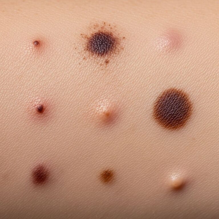
Trichofolliculoma is an uncommon small benign tumour originating from the hair follicle tissue. It typically presents as a small, solitary flesh-coloured or whitish papule or nodule, 2–6 mm in diameter, with a central pore from which fine vellus hairs protrude (like a small brush). Trichofolliculoma is sometimes called folliculoma.
What is trichofolliculoma?
Trichofolliculoma is a rare benign hair follicle hamartoma, classified as a hamartomatous neoplasm rather than a true neoplasm. It arises from the pilosebaceous unit and features a central dilated follicular infundibulum surrounded by immature hair follicles. First described by Miescher in 1944 and further characterized by Pinkus in 1965, it represents a malformation where pluripotent skin cells prematurely differentiate into hair structures.
These lesions develop spontaneously without association to genetic syndromes, sun exposure, or trauma in most cases. Rare congenital presentations exist, and exceptional links to amniotic band syndrome (ABS) have been reported due to developmental disruptions. Hamartomas like trichofolliculoma consist of well-developed cells in abnormal arrangements, forming cyst-like expansions of hair follicle openings.
Who gets trichofolliculoma?
Trichofolliculoma most commonly affects adults, with an average diagnosis age of 46 years (range 20–75 years). It shows no strong racial predilection and may slightly favour males, though evidence is limited. Lesions are typically solitary and sporadic, with rare multiple or congenital cases not tied to systemic conditions.
- Age: Predominantly adults; rare in children or congenital.
- Sex: Possible slight male predominance.
- Site: Face (especially nose: 40%), scalp, cheek (15%), ear (12%), neck, upper trunk; rarely lips, vulva, ear canal, genitalia.
Clinical features of trichofolliculoma
Apart from its physical appearance, trichofolliculoma shows no other signs or symptoms. It is usually asymptomatic, though rare painful or growing lesions occur, as in scalp cases with ABS. The classic presentation is a dome-shaped, skin-coloured papule or nodule (average 7 mm), often with a central umbilicated pore and tuft of fine, soft vellus hairs resembling a ‘troll doll’ or woolly wisp.
Not all lesions display hairs (only 12.2% in one study of 90 cases) or a visible pore (15.5%). Dermoscopy reveals characteristic signs:
- Erupting volcano pattern: Central dilated pore with tufted white hairs and radial vessels.
- Troll doll sign: Clusters of smooth white vellus hairs on a pink neoplasm.
- Firework pattern: Tan centre with radial dark brown projections, no pigment network.
In rare scalp variants, a well-defined whitish nodule (0.5 cm) with central umbilication and wool-like hairs may appear.
Diagnosis of trichofolliculoma
A small biopsy (punch or excisional under local anaesthetic) is the only definitive diagnosis for trichofolliculoma. Clinical features suggest it, but histology confirms and differentiates from mimics.
Histology of trichofolliculoma
Microscopically, trichofolliculoma shows a central dilated follicular infundibulum invaginating into the dermis, filled with keratin, hair shafts, and debris. The infundibulum base and sides feature radiating strands of hyperplastic epithelium forming immature secondary hair follicles. A fibrous stroma with mild inflammatory infiltrates surrounds it.
Key features include:
- Epidermal invagination with cystic dilatation.
- Hyperplastic infundibular epithelium with radiating trabeculae.
- Multiple abortive follicles opening into the central space.
- No atypia or malignancy.
Dermoscopy aids non-invasive assessment but biopsy rules out basal cell carcinoma or other adnexal tumours.
Differential diagnosis for trichofolliculoma
The histology of trichofolliculoma differentiates it from other benign follicular tumours with similar presentations. Common differentials include:
| Condition | Key Distinguishing Features |
|---|---|
| Dilated pore of Winer | Large, dilated pore with cornflake-like keratin plug; no secondary follicles; often on face/neck. |
| Pilar sheath acanthoma | Central pore with keratin; duct-like extensions; fewer radiating follicles. |
| Trichoepithelioma | Basaloid islands, fibrocystic stroma; no central infundibulum; may be multiple. |
| Trichoadenoma | Numerous uniform cysts; less organized follicles. |
| Basal cell carcinoma | Peripheral palisading, retraction artefact, atypia; requires biopsy exclusion. |
Rarely mimics fibrofolliculoma or sebaceous trichoma, but central hair tuft is distinctive for trichofolliculoma.
Management of trichofolliculoma
Observation suffices for asymptomatic lesions due to their benign nature—no malignant potential reported, though rare perineural invasion or recurrence post-excision exists. Complete surgical excision is curative for cosmesis, symptoms, or diagnostic confirmation, with low recurrence if margins clear.
- Conservative: No treatment needed; excellent prognosis.
- Surgical: Excision biopsy; preferred for facial lesions.
- Follow-up: Unnecessary unless recurrence suspected.
Laser ablation is unproven. Avoid unnecessary intervention.
Frequently Asked Questions (FAQs)
Is trichofolliculoma cancerous?
No, trichofolliculoma is a benign hamartoma with no reported malignant transformation.
Does trichofolliculoma go away on its own?
Typically stable; does not resolve spontaneously but requires no treatment.
Can trichofolliculoma appear anywhere on the body?
Primarily face (nose, cheek), scalp, ear; rarely lips, genitalia, ear canal.
How is trichofolliculoma diagnosed?
By skin biopsy showing central infundibulum with radiating follicles.
Is surgery always needed for trichofolliculoma?
No, only for cosmesis or confirmation; otherwise observe.
Related topics
- Dilated pore of Winer
- Trichoepithelioma
- Fibrofolliculoma
- Hair follicle tumours
- Adnexal tumours
References
- A unique presentation of trichofolliculoma in amniotic band syndrome — Journal of Surgical and Scientific Techniques in Dermatology. 2023. https://jsstd.org/a-unique-presentation-of-trichofolliculoma-in-amniotic-band-syndrome/
- Trichofolliculoma — MD Searchlight. 2024. https://mdsearchlight.com/skin-problems-and-treatments/trichofolliculoma/
- Trichofolliculoma — MalaCards (Monash University). 2025. https://www.malacards.org/card/trichofolliculoma
- Trichofolliculoma | Pore of Winer — DermNet NZ. 2024. https://dermnetnz.org/topics/trichofolliculoma
- Trichofolliculoma — VisualDx (Logical Images). 2025. https://www.visualdx.com/visualdx/diagnosis/trichofolliculoma?diagnosisId=52429&moduleId=21
Similar Articles


Read full bio of Sneha Tete