Trichomycosis Axillaris: Causes, Diagnosis & Treatment Guide
Superficial bacterial infection of axillary hair shafts causing coloured nodules, often asymptomatic but treatable with hygiene and antibiotics.

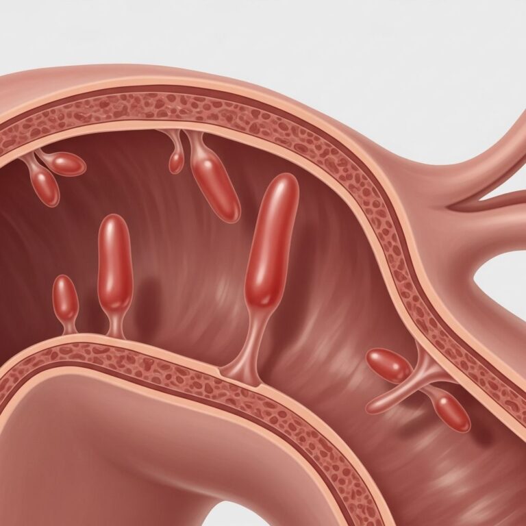
Trichomycosis axillaris is a superficial bacterial infection affecting axillary (underarm) hair shafts, characterised by the formation of yellow, black, or red granular nodules or concretions that adhere firmly to the hair. Despite its name suggesting a fungal origin, it is caused by bacterial overgrowth, primarily Corynebacterium species such as C. flavescens, C. tenuis, and C. propinquum, with occasional involvement of Serratia marcescens. This condition is benign, often asymptomatic, and prevalent in warm, moist environments, predominantly affecting young adult males due to higher apocrine sweat gland activity and poor hygiene practices. The concretions consist of densely packed bacteria embedded in a glycocalyx matrix, altering hair texture and sometimes causing odour. Recognition is crucial to differentiate it from more serious conditions, and treatment is straightforward, yielding excellent outcomes with minimal intervention.
What is trichomycosis axillaris?
Trichomycosis axillaris represents a chronic, superficial colonisation rather than invasive infection, where bacteria proliferate along the hair shaft in moist, occluded areas like the axillae. The term ‘trichomycosis’ is a misnomer, as histopathological examination reveals no fungal elements—only bacterial sheaths confirmed by Gram staining (Gram-positive rods) or culture. It forms part of the ‘corynebacterial triad’ alongside erythrasma (intertriginous coral-red fluorescence) and pitted keratolysis (plantar pits), often co-occurring in individuals with hyperhidrosis. Globally underreported due to its asymptomatic nature, prevalence can reach up to 28% in tropical regions among manual labourers, but it remains rare in temperate climates. The infection thrives in the alkaline environment created by apocrine sweat bacterial degradation, producing volatile fatty acids responsible for bromhidrosis.
Who gets trichomycosis axillaris?
Trichomycosis axillaris predominantly affects post-pubertal males, with studies reporting up to 97.4% male involvement, attributed to denser axillary hair and greater apocrine sweating. Age distribution peaks in young adults (20-40 years), correlating with active lifestyles, sports participation, and occupational exposure to heat/humidity. Risk escalates in obese individuals, those with hyperhidrosis, and poor personal hygiene, as moisture retention favours bacterial adhesion. Geographic predisposition favours tropical/subtropical climates (e.g., Latin America, Southeast Asia), where humidity exceeds 70%. Though axillary involvement is classic (nearly 100% of cases), ectopic sites include pubic hair (trichomycosis pubis), scrotum, intergluteal region, and rarely scalp or eyebrows via autoinoculation. Childhood cases are exceptional, typically scalp-localised. No racial predilection is noted, but socioeconomic factors like crowded living exacerbate transmission.
Causes
The primary aetiological agents are aerobic, Gram-positive Corynebacterium spp., with C. flavescens identified in 98% of cultured cases, forming yellow concretions; C. tenuis (white), C. propinquum (red), and C. otiidis (black) variants produce colour-specific nodules. These diphtheroids overgrow in the keratinaceous layer surrounding the hair shaft, forming adherent biofilms resistant to mechanical removal. Predisposing factors include:
- Poor hygiene: Inadequate washing allows bacterial proliferation in sweat-soaked hair.
- Hyperhidrosis: Excessive apocrine sweating provides nutrients and moisture.
- Occlusion: Tight clothing or axillary friction traps humidity.
- Obesity: Skin folds enhance moisture retention.
- Hot/humid climates: Optimal for bacterial growth (>30°C, high RH).
Transmission is likely autoinoculable or via fomites, not person-to-person.
Clinical features
Patients are often unaware of trichomycosis axillaris due to its subtlety, discovered incidentally during hygiene routines. Key signs include:
- Asymptomatic, soft, cylindrical concretions (1-2 mm thick) encasing hair shafts from base to tip, sparing the follicle/skin.
- Colour variants: yellow (most common, 60-80%), white, red, or black nodules, non-fluorescent under Wood’s lamp.
- Textural change: Hair feels sticky, matted, or ‘cemented,’ with increased thickness.
- Bromhidrosis: Foul, rancid odour from bacterial sweat metabolism (38-44% cases).
- Secondary features: Mild pruritus (1%), chromhidrosis (3%), sweat staining on clothes.
No inflammation, alopecia, or lymphadenopathy occurs. Pubic involvement mimics trichomycosis pubis; rare multi-site cases reported.
Complications
Trichomycosis axillaris is entirely benign with no systemic sequelae, scarring, or malignant potential. Rare issues include:
- Social embarrassment from odour/staining.
- Recurrent episodes if untreated (up to 50% without antibiotics).
- Co-infection with triad conditions (erythrasma 20-30%).
Untreated persistence is common but harmless.
Diagnosis
Diagnosis is clinical, based on characteristic nodules on axillary hair. Confirmation via:
- Microscopy: KOH prep shows Gram-positive branching rods in sheath; no hyphae.
- Culture: Selective media yield Corynebacterium (yellow colonies for C. flavescens).
- Wood’s lamp: Negative (distinguishes from erythrasma’s coral-red).
Biopsy rarely needed.
Differential diagnosis
| Condition | Key Differentiators |
|---|---|
| Pubic lice (pediculosis) | Mobile nits/eggs at follicle; itching; no concretions. |
| Trichosporon asahii (white piedra) | Soft, white nodules; fungal hyphae on KOH; tropical. |
| Chromhidrosis | Coloured sweat (not hair-bound); no nodules. |
| Pseudopelade/Scarring alopecia | Follicular destruction; bald patches. |
| Deodorant residue/stones | Washes off easily; no bacteria. |
Treatment
First-line: Shave/clip affected hair to eliminate bacterial reservoir (resolves 80-90%). Adjunctive topical antibacterials (apply BID x 1-2 weeks):
- Clindamycin 1% lotion (most common, 90% efficacy).
- Erythromycin 2% gel.
- Fusidic acid 2% cream (axillary preferred).
- Benzoyl peroxide 5% (alternative, irritant risk).
- Clotrimazole powder (empiric antifungal crossover).
For hyperhidrosis: Antiperspirants, botox, or iontophoresis. Systemic antibiotics (erythromycin) rarely for recalcitrant cases. Recurrence drops to <10% with combined therapy.
Prevention
- Daily axillary hygiene with antibacterial soap.
- Shaving/trimming during active infection.
- Breathable clothing; antiperspirants.
- Avoid occlusive environments.
Frequently asked questions
Is trichomycosis axillaris contagious?
No, it spreads via poor hygiene/autoinoculation, not direct contact.
Does it go away on its own?
Often persists indefinitely without intervention.
Can it affect pubic hair?
Yes, termed trichomycosis pubis; similar management.
Is it dangerous?
Benign; no scarring or systemic risks.
How long until treatment works?
Resolution in 7-14 days with adherence.
References
- An Overview of Trichobacteriosis (Trichomycosis) — National Library of Medicine (PMC). 2023-10-01. https://pmc.ncbi.nlm.nih.gov/articles/PMC10600505/
- Doctor my armpit hair has turned yellow — Royal Australian College of General Practitioners (RACGP). 2018-09-01. https://www1.racgp.org.au/ajgp/2018/september/doctor-my-armpit-hair-has-turned-yellow
- Trichomycosis axillaris — DermNet NZ (Authoritative dermatology resource). 2024-01-01. https://dermnetnz.org/topics/trichomycosis-axillaris
- Trichomycosis: Definition, treatment, symptoms, and causes — Medical News Today. 2023-05-15. https://www.medicalnewstoday.com/articles/trichomycosis
- Trichomycosis: Causes, Symptoms, and Treatment — Healthline. 2023-08-20. https://www.healthline.com/health/trichomycosis
Similar Articles


Read full bio of medha deb