Cystoscopy: What To Expect, Risks, And Recovery Guide
Explore the essential diagnostic tool for bladder and urethra health, from preparation to recovery and beyond.

Cystoscopy serves as a cornerstone procedure in urology, enabling healthcare providers to directly visualize the interior of the bladder and urethra. This minimally invasive technique uses a specialized instrument called a cystoscopea thin tube equipped with a light and camerato identify abnormalities such as tumors, stones, infections, or structural issues. By offering real-time imaging, it facilitates accurate diagnosis and often immediate treatment, reducing the need for more extensive surgeries.
The Anatomy Behind the Procedure
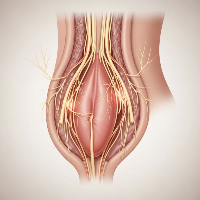
Before delving into the procedure itself, it’s helpful to understand the relevant anatomy. The urethra is the tube that carries urine from the bladder out of the body, while the bladder acts as a storage organ for urine. In men, the urethra is longer, passing through the prostate and penis; in women, it is shorter. Conditions affecting these structures can lead to symptoms like blood in urine, frequent infections, pain during urination, or difficulty emptying the bladder. Cystoscopy provides unparalleled access to inspect these areas directly.
Primary Reasons for Recommending Cystoscopy
Healthcare professionals recommend cystoscopy for a variety of diagnostic and therapeutic purposes. Key indications include:
- Investigating hematuria (blood in the urine), which could signal cancer, stones, or infections.
- Diagnosing recurrent urinary tract infections (UTIs) to identify underlying causes like bladder stones or diverticula.
- Evaluating urinary retention or obstruction due to strictures, enlarged prostate, or tumors.
- Detecting bladder or urethral cancer, polyps, or abnormal growths.
- Assessing pain or discomfort during urination (dysuria) without obvious external causes.
- Monitoring known conditions, such as post-treatment surveillance for bladder cancer.
This procedure is particularly valuable because it allows for biopsiessmall tissue samples taken for microscopic analysisto confirm suspicions of malignancy or inflammation.
Types of Cystoscopes and Their Applications
There are two main types of cystoscopes: flexible and rigid, each suited to different scenarios.
| Type | Description | Typical Use | Anesthesia | Duration |
|---|---|---|---|---|
| Flexible | Thin, bendable tube with camera | Diagnostic exams, office-based | Local (gel) | 10-15 minutes |
| Rigid | Straight, sturdier tube | Treatment, biopsies, stone removal | General or spinal | 20-30 minutes |
Flexible cystoscopes are preferred for initial diagnostics due to patient comfort and outpatient feasibility, while rigid ones enable more precise interventions.
Preparing for Your Cystoscopy
Preparation is straightforward but crucial for safety and comfort. Patients are typically advised to:
- Empty the bladder just before the procedure and possibly provide a urine sample to rule out active infections.
- Discuss medications, especially blood thinners like aspirin or anticoagulants, which may need temporary cessation.
- Arrange transportation if sedation or anesthesia is planned, as driving is unsafe post-procedure.
- Fast if general anesthesia is used, following specific guidelines from the care team.
Women may be advised to avoid vaginal products, and men should note any prostate issues. Always inform your doctor of allergies, implants, or heart conditions.
Step-by-Step: What Happens During a Flexible Cystoscopy
A flexible cystoscopy is often performed in an outpatient clinic. Here’s the typical sequence:
- Undress from the waist down and lie on an exam table, covered by a gown.
- A numbing gel is applied to the urethra to minimize discomfortno needles required.
- The lubricated cystoscope is gently inserted through the urethra into the bladder, which may cause mild pressure.
- Saline solution fills the bladder to expand it, improving visibility; patients often feel an urge to urinate.
- The urologist inspects the urethra and bladder walls via a monitor, rotating the scope for full coverage.
- If needed, tiny tools pass through the scope for biopsy or minor treatments.
- The scope is withdrawn smoothly, concluding the exam.
Most patients tolerate it well, with only brief stinging or fullness sensations.
Rigid Cystoscopy: When More Intervention Is Needed
For procedures requiring greater maneuverability, a rigid cystoscope under anesthesia is used. Performed in a hospital or surgical center, it follows similar steps but allows for advanced actions like:
- Removing bladder stones or small tumors.
- Treating strictures or injecting medications into the bladder wall.
- Performing larger biopsies or cauterizing bleeding sites.
Anesthesia ensures no pain, though recovery involves monitoring vital signs.
Safety Profile: Risks and Complications
While generally safe, cystoscopy carries minor risks, including:
- Temporary burning or blood in urine for 1-2 days.
- Infection (rare, prevented by antibiotics if needed).
- Urethral irritation or spasm, easing with time.
- Rarely, bladder perforation or heavy bleeding, requiring prompt care.
Success rates are high, with complications under 5% in experienced hands. Contact your doctor for fever, severe pain, or persistent bleeding post-procedure.
Aftercare and Recovery Expectations
Recovery is quick for office-based flexible scopesyou can resume normal activities soon after, drinking plenty of fluids to flush the system. For rigid procedures under anesthesia:
- Rest for 24 hours; avoid strenuous activity.
- Expect pink-tinged urine for a few days; use over-the-counter pain relief for discomfort.
- Avoid heavy lifting or intercourse for 48 hours.
- Follow-up discusses findings; biopsy results take 3-7 days.
Hydration is key to prevent clots and promote healing.
Patient Experiences and Tips
Many describe the procedure as more tolerable than anticipated, likening it to a prolonged urination sensation. Tips include:
- Breathe deeply and relax muscles during insertion.
- Request to watch the screen for distraction and education.
- Wear loose clothing post-procedure for comfort.
Open communication with your urologist alleviates anxiety.
Advancements and Alternatives
Modern innovations include narrower scopes for less discomfort and blue-light cystoscopy for enhanced cancer detection using fluorescent dyes. Alternatives like ultrasound or CT scans offer non-invasive views but lack the precision of direct visualization. For certain cases, ureteroscopy extends inspection to the kidneys.
Frequently Asked Questions (FAQs)
Is cystoscopy painful?
Local anesthesia minimizes pain to mild discomfort or pressure. Sedation makes it painless for most.
How long do results take?
Immediate visual findings; biopsies analyzed in days to a week.
Can women have cystoscopy during pregnancy?
Usually avoided unless essential; discuss risks with your doctor.
Does insurance cover it?
Typically yes, as a standard diagnostic test—verify with your provider.
What’s the difference from a catheter?
Cystoscopy includes imaging and tools; catheters only drain urine.
Cystoscopy empowers precise urological care, often resolving mysteries behind urinary symptoms swiftly and effectively.
References
- How a cystoscopy is done – NHS NHS. 2023. https://www.nhs.uk/tests-and-treatments/cystoscopy/how-its-done/
- Cystoscopy UCSF Benioff Children’s Hospitals. 2024. https://www.ucsfbenioffchildrens.org/medical-tests/cystoscopy
- Cystoscopy & Ureteroscopy NIDDK. 2023-10-23. https://www.niddk.nih.gov/health-information/diagnostic-tests/cystoscopy-ureteroscopy
- Cystoscopy for Men University of Rochester Medical Center. 2024. https://www.urmc.rochester.edu/encyclopedia/Content?contentTypeID=92&ContentID=P07771
- Cystoscopy Procedure Advocate Health Care. 2024. https://www.advocatehealth.com/health-services/urology/services-treatments/cystoscopy
- Cystoscopy (Bladder Endoscopy) YouTube (Medical Animation). 2018. https://www.youtube.com/watch?v=iLeqYPJyG_A
- Cystoscopy: Purpose, Procedure, Risks & Recovery Cleveland Clinic. 2024-01-29. https://my.clevelandclinic.org/health/diagnostics/16553-cystoscopy
Similar Articles


Read full bio of Sneha Tete