Iris Hypoplasia: Causes, Symptoms, And Treatment Guide
Explore the causes, symptoms, diagnosis, and management strategies for iris hypoplasia, a congenital eye condition affecting iris development.

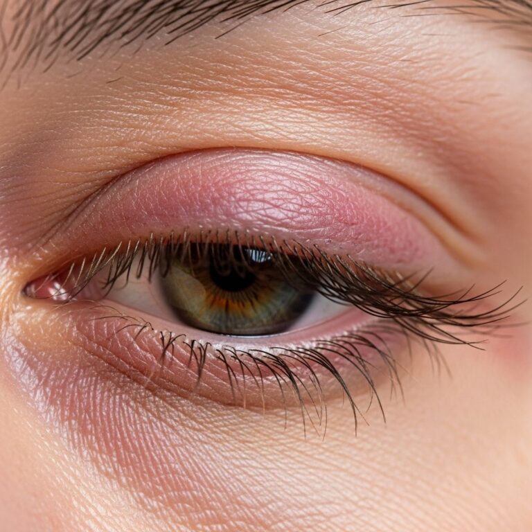
Iris hypoplasia refers to the underdevelopment or incomplete formation of the iris, the colored part of the eye that controls light entry by adjusting the pupil size. This congenital condition arises during fetal eye development and can range from mild underdevelopment to near-total absence of iris tissue, impacting visual function significantly.
The Anatomy and Role of the Iris
The iris is a circular, pigmented diaphragm surrounding the pupil. It contains two muscle layers: the dilator muscle, which widens the pupil in dim light, and the sphincter muscle, which narrows it in bright conditions. These muscles enable photophobia reduction and focus adjustment. In iris hypoplasia, insufficient tissue formation disrupts these functions, leading to excessive light entry and poor vision quality.
Normal iris development begins around the 5th week of gestation, involving neural crest cells and genetic signaling pathways. Disruptions here result in hypoplastic irises, often detectable at birth through enlarged or irregular pupils.
Types and Spectrum of Iris Hypoplasia
Iris hypoplasia manifests in varying degrees:
- Mild hypoplasia: Subtle thinning of iris tissue with minimal pupil irregularity.
- Moderate hypoplasia: Partial iris absence, creating keyhole-shaped pupils.
- Severe forms (aniridia): Near-complete iris absence, exposing large pupils.
Isolated hypoplasia differs from syndromic cases, such as congenital aphakia-iris hypoplasia-microphthalmia-microcornea syndrome, involving small eyes, absent lenses, and additional anomalies like nystagmus and glaucoma.
Genetic and Developmental Causes
Most cases stem from genetic mutations affecting eye ontogeny. The PAX6 gene, crucial for ocular structure formation, is a primary culprit. Mutations yield nonfunctional proteins, halting iris differentiation during embryogenesis.
Other implicated genes regulate neural crest migration and anterior segment development. Inheritance patterns include autosomal dominant (common in aniridia) and sporadic mutations. Environmental factors like maternal diabetes rarely contribute, but genetics predominate.
Recognizing Symptoms and Clinical Signs
Symptoms often appear at birth or infancy:
- Photophobia: Intense light discomfort due to poor pupil constriction.
- Reduced visual acuity: Blurred vision from light scatter.
- Nystagmus: Involuntary eye oscillations.
- Strabismus: Eye misalignment.
- Glare and halos around lights.
Physical signs include oversized, eccentric, or oddly shaped pupils. In severe cases, leukocoria (white pupil reflex) signals associated issues like persistent fetal vasculature. Long-term, patients risk glaucoma (elevated intraocular pressure), cataracts, and foveal hypoplasia (poor central vision).
Associated Conditions and Complications
| Condition | Description | Prevalence in Hypoplasia |
|---|---|---|
| Glaucoma | Pressure buildup damaging optic nerve | Common in adolescence |
| Cataracts | Lens clouding | 50-85% of cases |
| Foveal Hypoplasia | Underdeveloped macula | Frequent, impairs acuity |
| Optic Nerve Hypoplasia | Underdeveloped nerve | 10% in severe forms |
| Microphthalmia/Microcornea | Small eye/cornea | Syndromic variants |
Systemic links are rare but include olfactory deficits or developmental delays in PAX6-related aniridia. Regular monitoring prevents vision-threatening complications.
Diagnostic Approaches
Diagnosis combines clinical exam and imaging:
- Slit-lamp biomicroscopy: Reveals iris thinning or absence.
- Fundoscopy: Assesses retina, fovea, optic nerve.
- Ultrasound biomicroscopy (UBM): High-res imaging of anterior segment.
- Optical coherence tomography (OCT): Detailed foveal analysis.
- Genetic testing: Confirms PAX6 or related mutations.
Newborn screening via red reflex tests flags leukocoria. Early diagnosis enables proactive care.
Treatment and Management Strategies
No cure exists for missing iris tissue, but interventions mitigate effects:
- Tinted contact lenses or iris prosthetic devices: Reduce light entry, improve cosmesis.
- Pharmacologic agents: Miotics constrict pupils; glaucoma drops manage pressure.
- Surgical options: Iris reconstruction for select cases; cataract/glaucoma procedures as needed.
- Low-vision aids: Magnifiers, filters for daily function.
Multidisciplinary care involves ophthalmologists, geneticists, and low-vision specialists. Lifelong surveillance tracks complications.
Living with Iris Hypoplasia: Daily Life and Adaptation
Affected individuals adapt via environmental modifications: polarized sunglasses, hats, dimmable lighting. School/work accommodations include preferential seating and assistive tech.
Psychosocial support addresses self-esteem issues from visible anomalies. Many lead fulfilling lives with proper management, pursuing education and careers despite reduced acuity.
Prognosis and Long-Term Outlook
Prognosis varies: mild cases yield near-normal vision; severe aniridia risks progressive loss from glaucoma/cataracts. Early intervention preserves function. Genetic counseling aids family planning, especially with dominant inheritance.
Research Advances and Future Directions
Ongoing studies explore gene therapy targeting PAX6, stem cell iris regeneration, and advanced prosthetics. Clinical trials assess novel implants for light regulation. Patient registries enhance understanding of rare variants.
Frequently Asked Questions (FAQs)
What causes iris hypoplasia?
Primarily genetic mutations like those in the PAX6 gene disrupt fetal iris development.
Is iris hypoplasia the same as aniridia?
Aniridia is a severe form with near-total iris absence; hypoplasia encompasses milder underdevelopment.
Can iris hypoplasia be treated surgically?
Surgery improves appearance and function in some cases but doesn’t restore natural tissue.
Does it affect both eyes?
Typically bilateral and symmetric, though asymmetry occurs.
What is the inheritance pattern?
Often autosomal dominant, but sporadic cases are common.
This guide empowers patients and families with knowledge for informed decisions. Consult eye specialists for personalized advice.
References
- Congenital aphakia-iris hypoplasia-microphthalmia-microcornea syndrome — Orphanet. 2023. https://www.orpha.net/en/disease/detail/617449
- Aniridia – Genetics — MedlinePlus Genetics (U.S. National Library of Medicine). 2024-02-15. https://medlineplus.gov/genetics/condition/aniridia/
- Aniridia — Minnesota Department of Health. 2023. https://www.health.state.mn.us/diseases/cy/aniridia.html
- Coloboma: Types, Causes & Associated Conditions — Cleveland Clinic. 2024-01-10. https://my.clevelandclinic.org/health/diseases/22682-coloboma
- Hypoplasia of the iris (Concept Id: C0344539) — NCBI MedGen. 2022. https://www.ncbi.nlm.nih.gov/medgen/91029
Similar Articles


Read full bio of Sneha Tete