Vascular Birthmarks: Types, Causes, and Treatment Options
Comprehensive guide to understanding vascular birthmarks, their causes, symptoms, and modern treatment approaches.

Understanding Vascular Birthmarks
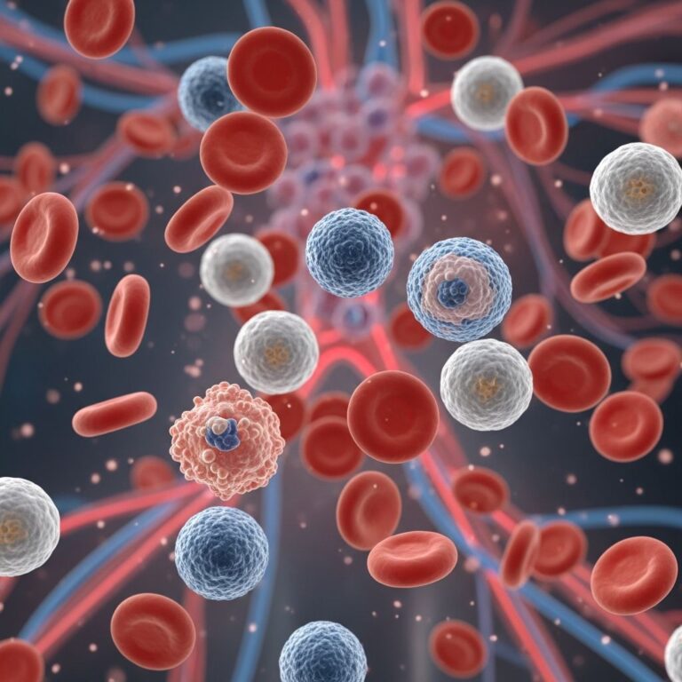
Vascular birthmarks are skin abnormalities that result from the abnormal development of blood vessels, including arteries, veins, lymphatic vessels, or capillaries. These birthmarks represent one of the most common types of skin conditions present at birth or appearing within the first weeks and months of life. Understanding the different types of vascular birthmarks, their underlying causes, and available treatment options is essential for parents and individuals seeking to make informed decisions about their health care.
Vascular birthmarks fall into two major categories: hemangiomas and vascular malformations. While these conditions may appear similar on the surface, they differ significantly in their development, progression, and treatment requirements. Hemangiomas are benign tumors composed of rapidly proliferating endothelial cells, whereas vascular malformations represent structural abnormalities in blood vessel formation that persist throughout life.
Types of Vascular Birthmarks
Hemangiomas
Hemangiomas are the most common type of vascular birthmark, affecting approximately 7 percent of the population, with a higher prevalence in Caucasian children, premature infants, and females. These benign growths consist of clusters of abnormal blood vessel cells that typically appear within the first few days to weeks of an infant’s life.
The initial presentation of hemangiomas often resembles a small red scratch or tiny bump on the skin. During the early stage, known as the proliferative phase, hemangiomas grow rapidly for approximately six months to one year. This rapid growth phase is a natural part of the hemangioma’s development and does not necessarily indicate a problem requiring immediate intervention.
One of the most encouraging aspects of hemangiomas is their natural history of resolution. Research demonstrates that approximately 50 percent of hemangiomas spontaneously disappear by age 5, and approximately 90 percent resolve completely by age 9 without any medical treatment. This natural regression occurs as the growth phase transitions into an involution phase, during which the hemangioma gradually loses its red coloration and progressively shrinks.
Vascular Malformations
Unlike hemangiomas, vascular malformations are permanent structural abnormalities of blood vessels that develop during fetal life. These malformations do not spontaneously regress and continue to grow proportionally as children grow. The classification of vascular malformations depends on the specific type of blood vessel involved in the abnormality.
Port-Wine Stains (Capillary Malformations)
Port-wine stains, also known as capillary malformations, are flat, reddish or purplish birthmarks that are typically visible at birth. These lesions result from enlarged capillaries in the skin and are frequently located on the face, head, and neck, though they can appear anywhere on the body. Port-wine stains develop due to a deficiency in the nerve fibers that normally regulate blood vessel constriction in the affected area.
An important characteristic of port-wine stains is their progressive nature. As children grow, the skin surrounding the port-wine stain often thickens, and the discoloration becomes increasingly noticeable and darker. The lesions typically darken from a light pink color to deeper shades of purple and red as the child ages. While port-wine stains are usually not dangerous, they generally do not fade on their own and may warrant evaluation by a vascular birthmark specialist to rule out associated conditions.
Venous Malformations
Venous malformations represent abnormalities involving deep veins and typically appear as bluish marks on the skin. These malformations consist of tangled, enlarged veins lacking proper smooth muscle along their vessel walls, which causes them to enlarge and rupture easily. Although venous malformations are usually present at birth, some may not become visibly apparent until years later.
Venous malformations commonly occur on the jaw, cheek, tongue, and lips. These lesions have low blood flow and feel soft to the touch. A distinctive characteristic is that the color of the lesion may disappear when compressed, a useful diagnostic feature. Some venous malformations can be managed through injection therapy or sclerosis, a procedure that helps reduce the size and appearance of the abnormal veins.
Lymphatic Malformations
Lymphatic malformations develop when the fluid in lymphatic vessels does not drain properly, causing excess fluid to accumulate and create cysts or swollen, enlarged vessels. These malformations most commonly occur in the head and neck region, though they can develop anywhere in the body. When lymphatic malformations occur close to the skin’s surface, they may resemble bruises.
Large cystic masses in the neck are known as cystic hygromas and represent a specific type of lymphatic malformation. These conditions can interrupt the normal pathway of the lymph system, causing the vessels to widen and leading to swelling. In some cases, the enlarging vessels can allow blood to enter them, further complicating the clinical picture. Lymphatic malformations affecting the skin can cause recurrent skin infections, scabbing, and weeping from blisters. These lesions are particularly difficult to treat, though newer injection therapies show promise.
Arteriovenous Malformations
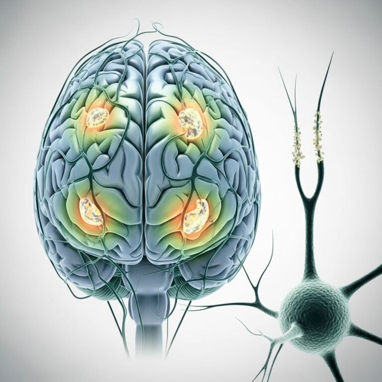
Arteriovenous malformations (AVMs) represent abnormal direct connections between arteries and veins, bypassing the normal capillary bed. These high-flow lesions are believed to result from improper formation of the artery-capillary-vein connections during early pregnancy. AVMs may affect a specific localized area or be more widespread throughout the body.
AVMs commonly appear on the trunk, arms, and legs. In childhood, the pink or red coloration associated with an AVM can be mistaken for a hemangioma. However, due to the high flow of blood through the abnormal connection, AVMs become increasingly noticeable over time. As AVMs progress, the skin may become darker red or purple, nearby veins may enlarge, a palpable pulse may be felt in the affected area, and the region may feel warm to the touch. When AVMs occur within the brain, specialized neurosurgical intervention is required.
Macular Stains and Salmon Patches
Macular stains, also known as salmon patches, are characterized by pink to red marks that may appear anywhere on the body. The most common types of these temporary vascular birthmarks are angel’s kisses and stork bites. Angel’s kisses appear as marks on the forehead, nose, upper lip, and eyelids and typically disappear with age. Stork bites are marks located on the back of the neck that also usually fade as the child grows.
Causes of Vascular Birthmarks
Hemangioma Etiology
The exact cause of hemangiomas remains incompletely understood despite extensive research. Hemangiomas result when endothelial cells migrate too rapidly during fetal development, creating widened capillaries around the developing tumor. This excessive cell migration increases both the size and appearance of the hemangioma. Current research indicates some potential genetic links, though these findings remain inconclusive.
Vascular Malformation Etiology
Vascular malformations occur when blood vessels develop abnormally during fetal life. For port-wine stains specifically, the affected area lacks the small nerve fibers necessary to narrow and regulate the vessels properly. Telangiectatic nevi, another type of vascular malformation, develop due to how certain blood vessels form during pregnancy. Unlike hemangiomas, there is no known genetic cause for vascular malformations, and current understanding suggests they result from developmental abnormalities during embryogenesis.
Clinical Features and Symptoms
Vascular birthmarks present with varied clinical features depending on their type and severity. Hemangiomas typically appear as red bumps or patches that may be slightly raised or flat. Some hemangiomas may have overlying skin that appears bruised or discolored.
Vascular malformations can cause pain, swelling, and discomfort due to engorgement of the abnormal vessels. In some cases, underlying bone and soft tissues may be stimulated to grow abnormally fast in relation to surrounding tissues, leading to asymmetry or functional impairment. Arteriovenous malformations can redirect blood flow away from tissues downstream of the malformation, leading to poor oxygen supply and potential complications. The specific symptoms depend on the location, type, and extent of the vascular abnormality.
Diagnosis and Evaluation
Most vascular birthmarks can be diagnosed through careful physical examination by a healthcare provider. The appearance, location, and characteristics of the lesion often provide sufficient information for diagnosis. For complex cases or when deeper involvement is suspected, imaging studies such as ultrasound, magnetic resonance imaging (MRI), or computed tomography (CT) scans may be necessary to fully characterize the extent of the vascular abnormality and its relationship to surrounding structures.
Multidisciplinary evaluation by specialists in dermatology, pediatric surgery, plastic surgery, interventional radiology, and otolaryngology can provide comprehensive assessment and coordinated treatment planning. This team approach ensures that all aspects of the patient’s condition are considered when formulating a treatment strategy.
Treatment Options
Observation and Watchful Waiting
Given the high rate of spontaneous resolution in hemangiomas, many lesions benefit from a conservative approach of observation and monitoring. Regular follow-up appointments with a healthcare provider allow tracking of the hemangioma’s natural history. Parents and caregivers receive education about expected progression and warning signs that might warrant intervention.
Laser Therapy
Laser therapy has emerged as one of the most effective treatments for certain vascular birthmarks, particularly port-wine stains and superficial hemangiomas. Pulsed dye lasers work by targeting the hemoglobin in red blood cells, causing selective destruction of abnormal blood vessels while minimizing damage to surrounding tissue. Laser therapy can be particularly helpful in children to lighten the appearance of lesions and prevent progressive darkening.
Pharmacological Treatments
Systemic medications may be prescribed for hemangiomas that are growing rapidly, interfering with vital functions, or causing significant disfigurement. These medications work by reducing the proliferative activity of the hemangioma cells and promoting involution of the lesion.
Injection Therapy and Sclerosis
Certain vascular malformations, particularly venous malformations, may respond well to injection therapy or sclerotherapy. These procedures involve injecting sclerosing agents directly into the abnormal vessels to cause them to collapse and be resorbed by the body. This approach can significantly reduce the size and appearance of accessible lesions.
Surgical Intervention
Surgical removal may be considered for hemangiomas that are interfering with vital functions such as vision or breathing, for lesions that have become ulcerated and are not responding to other treatments, or for certain vascular malformations that have not responded to less invasive approaches. Modern surgical techniques aim to minimize scarring while effectively removing the abnormal vascular tissue.
Interventional Radiology
Interventional radiologists can perform minimally invasive procedures to treat vascular malformations. Techniques such as embolization can reduce blood flow through arteriovenous malformations and other high-flow lesions, helping to control symptoms and prevent progression.
Prognosis and Long-Term Outcomes
The prognosis for vascular birthmarks varies considerably depending on the type and severity of the lesion. Hemangiomas have an excellent long-term prognosis, with the vast majority resolving spontaneously during childhood without requiring treatment. Port-wine stains do not fade spontaneously but respond well to laser therapy when treatment is initiated early. Vascular malformations require ongoing management, as they continue to grow with the child and may develop complications requiring intervention.
Frequently Asked Questions
Q: Are all birthmarks dangerous?
Most vascular birthmarks are benign and not dangerous, though some may require monitoring or treatment for cosmetic reasons or to prevent complications. Certain lesions, such as those interfering with vision or breathing, warrant medical evaluation and possible intervention.
Q: Will my child’s hemangioma go away on its own?
Yes, approximately 90 percent of hemangiomas resolve completely by age 9 without treatment. However, regular monitoring by a healthcare provider is important to ensure normal progression and to identify any complications early.
Q: Can vascular malformations be cured?
Vascular malformations are permanent structural abnormalities that do not cure spontaneously. However, various treatment options can help manage symptoms, improve appearance, and prevent complications. Treatment decisions should be individualized based on the type, location, and extent of the malformation.
Q: What is the best treatment for port-wine stains?
Laser therapy is currently the most effective treatment for port-wine stains, particularly when initiated early in childhood. Multiple treatment sessions may be necessary to achieve optimal lightening of the lesion.
Q: Should I be concerned about my infant’s hemangioma?
Most infantile hemangiomas are not a cause for concern and will resolve naturally. However, hemangiomas that grow rapidly, interfere with vital functions, become ulcerated, or are located near sensitive areas should be evaluated by a healthcare provider to determine if treatment is necessary.
References
- Birthmarks and Vascular Anomalies — Gillette Children’s Specialty Healthcare. 2024. https://www.gillettechildrens.org/conditions-care/birthmarks-and-vascular-anomalies
- About Vascular Birthmarks — South Tees Hospitals NHS Foundation Trust. 2024. https://www.southtees.nhs.uk/services/plastic-surgery/vascular-birthmark-clinic/about-vascular-birthmarks/
- Vascular Birthmarks — Center for Craniofacial Care, Children’s Hospital of Richmond. 2024. https://www.chrichmond.org/services/center-for-craniofacial-care/conditions-treated/vascular-birthmarks/
- What is a Vascular Birthmark? — Pediatric Cleft and Craniofacial Center, University of Rochester Medical Center. 2024. https://www.urmc.rochester.edu/childrens-hospital/craniofacial/vascular-birthmark.aspx
- Vascular Birthmarks — St. Louis Children’s Hospital. 2024. https://www.stlouischildrens.org/conditions-treatments/vascular-birthmarks
- Birthmark Causes & Treatments — Cleveland Clinic. 2024. https://my.clevelandclinic.org/health/diseases/12159-birthmarks
- Birthmark | Mole | Hemangioma | Mongolian Spot — MedlinePlus, National Library of Medicine. 2024. https://medlineplus.gov/birthmarks.html
Similar Articles


Read full bio of medha deb