Vascular Malformations with Steal Phenomena
Exploring vascular malformations that cause steal phenomena, their cutaneous signs, syndromes, and management strategies in dermatology.

Vascular malformations (VMs) are congenital anomalies of blood and lymphatic vessels that can lead to significant clinical complications, including the
steal phenomenon
. The steal phenomenon occurs when high-flow shunting through malformed vessels diverts blood away from surrounding normal tissues, causing ischemia, pallor, ulceration, and pain. This article examines key types of VMs associated with steal phenomena, their dermatological manifestations, associated syndromes, diagnostic approaches, and management strategies.What is the Steal Phenomenon?
The
steal phenomenon
, also known as vascular steal syndrome, refers to reduced perfusion in tissues adjacent to or distal to a high-flow vascular malformation, such as an arteriovenous malformation (AVM). Blood is shunted through low-resistance anomalous channels, depriving normal capillary beds of oxygenated blood. Clinically, this manifests as pallor, coolness, paresthesia, cyanosis, rest pain, and ulceration in affected areas. In cutaneous presentations, a characteristic white halo of vasoconstriction surrounds telangiectatic lesions, indicating local ischemia.Histologically, steal-related changes include endothelial hyperplasia and reactive angioproliferation driven by hypoxia and elevated venous pressures. Deep AVMs may remain occult until hemorrhage or necrosis reveals them.
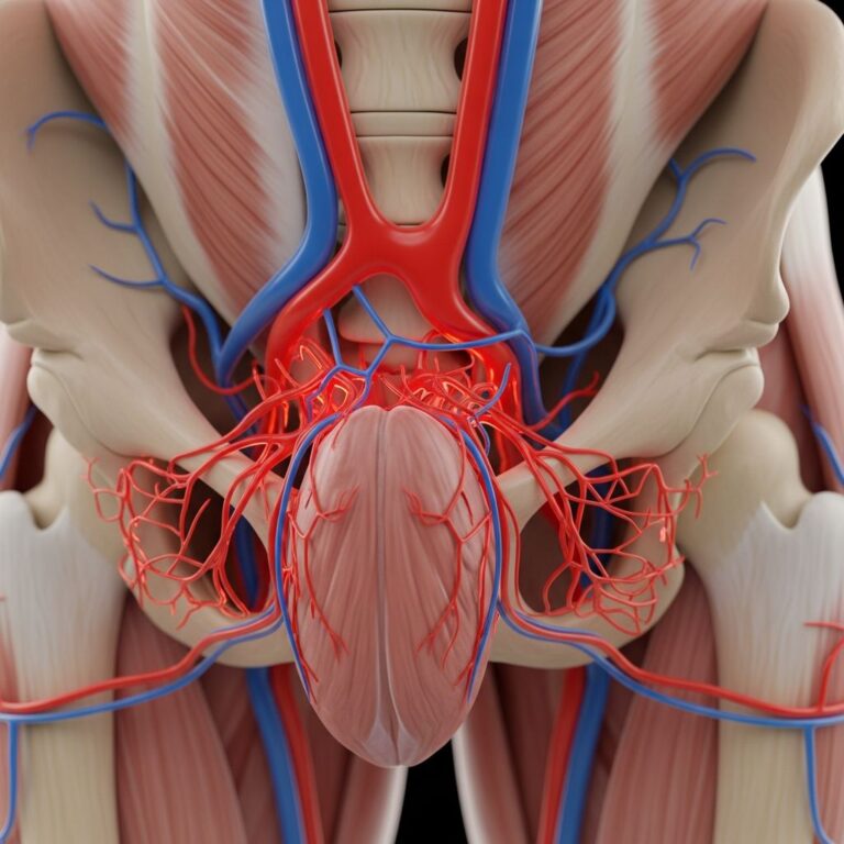
Arteriovenous Malformations (AVMs)
**Arteriovenous malformations** are high-flow VMs connecting arteries directly to veins, bypassing capillaries. They often present as warm, pulsatile masses with overlying skin changes due to steal.
- Cutaneous steal signs: Pallor surrounding fine telangiectasias or combined venous-lymphatic malformations overlying subcutaneous AVMs (Fig. 1a-b).
- Mature AVMs: Enlarged, pulsating subcutaneous masses with soft tissue hypertrophy, fibrosis, and warmth.
- Complications: Distal ischemia, necrosis, ulceration, and acroangiodermatitis—a reactive hyperplasia mimicking Kaposi sarcoma with violaceous plaques on extensor surfaces.
Acroangiodermatitis arises from chronic venous hypertension, presenting as brown macules, papules, and nodules that ulcerate, predominantly on dorsal feet and toes. Doppler ultrasound confirms high-flow shunting.
Capillary Malformation-Arteriovenous Malformation (CM-AVM) Syndrome
**CM-AVM syndrome** is an autosomal dominant disorder caused by RASA1 or EPHB4 mutations, featuring multiple small capillary malformations (CMs) associated with underlying AVMs.
Characteristic skin lesions are multifocal, acquired, round-to-oval pink-red macules or papules (<1-3 cm), often with a
white halo
indicating vascular steal. These differ from classic port-wine stains by their small size, multiplicity, and arterial Doppler flow.- Associated AVMs: High-flow lesions in skin, muscle, brain, spine, or bone, risking hemorrhage or heart failure.
- Diagnosis: Clinical suspicion plus imaging (MRI/MRA, ultrasound); genetic testing confirms.
Lesions may exhibit hypotrichosis or brownish hues; vascular blebs appear in adults.
Other Capillary Malformations and Steal
Port-wine stains (PWS), the most common CMs (0.3% newborns), are slow-flow ectatic capillaries in the dermis. While typically not high-flow, certain PWS show steal-like halos due to autonomic dysregulation.
In
Sturge-Weber syndrome (SWS)
, facial PWS in V1 trigeminal distribution overlies leptomeningeal angiomas, causing cortical ischemia, calcification, glaucoma, and seizures. SWS results from somatic GNAQ mutations.Klippel-Trenaunay Syndrome (KTS) and Related Overgrowth Syndromes
**Klippel-Trenaunay syndrome** combines capillary-lymphatic-venous malformations with limb overgrowth.
| Syndrome | Key Features | Steal-Related Skin Changes |
|---|---|---|
| Klippel-Trenaunay (KTS) | Limb hypertrophy, venous varicosities, CMs, lymphatic anomalies | Venous eczema, hyperpigmentation, lipodermatosclerosis, ulceration |
| Servelle-Martorell | Venous malformations with bony hypotrophy, limb shortening | Chronic venous insufficiency (CVI) signs: atrophie blanche, calcification |
| Parkes Weber | Multiple AVMs with limb overgrowth | Distal ischemia, necrosis from steal |
Truncular venous malformations (VMs) cause CVI with eczema, pigmentation, and ulcers, exacerbated by steal in combined forms.
Venous and Glomuvenous Malformations
**Venous malformations** present with dilation, hypoplasia, or aneurysms leading to venous hypertension. Glomuvenous malformations (GVMs) show cobblestone, tender plaques due to glomus cells (GLMN mutations).
VMs with TEK/TIE2 mutations feature ectatic vessels lacking smooth muscle, prone to thrombosis and steal.
Clinical Presentation and Diagnosis
Skin findings vary by depth and flow:
- Superficial: Telangiectasias, pallor halos, acroangiodermatitis.
- Deep: Pulsatile masses, hypertrophy, occult until bleed.
- Syndromic: Multifocal CMs, overgrowth, CVI.
Diagnostic tools:
- Ultrasound/Doppler: Flow assessment.
- MRI/MRA: Extent mapping.
- Genetic testing: For syndromes.
- Biopsy: Rarely, for equivocal cases.
Differential Diagnosis
- Infantile hemangiomas (proliferate then involute).
- Pyogenic granuloma.
- Kaposi sarcoma (acroangiodermatitis mimic).
- Stasis dermatitis without malformation.
Management and Treatment
Treatment targets shunting and symptoms.
- Embolization/Sclerotherapy: First-line for AVMs to reduce flow.
- Surgery: Resection for localized lesions; amputation in severe steal necrosis.
- Laser (PDL): For CMs, though less effective in high-flow.
- Compression: For venous components.
- Sirolimus: Emerging for complex VMs.
Multidisciplinary care (dermatology, interventional radiology, genetics) is essential. Screening for visceral AVMs prevents complications.
Frequently Asked Questions (FAQs)
What causes the white halo around CM-AVM lesions?
The white halo represents vascular steal, where blood is shunted away from surrounding tissues into the high-flow malformation.
Can steal phenomena lead to amputation?
Yes, severe distal ischemia and necrosis from AVM steal can necessitate amputation, as seen in hand AVMs.
Is CM-AVM syndrome inherited?
Yes, autosomal dominant via RASA1 or EPHB4 mutations; family screening is recommended.
How is KTS distinguished from Parkes Weber?
KTS has slow-flow venous/lymphatic malformations with overgrowth; Parkes Weber features fast-flow AVMs.
What is the prognosis for untreated AVMs?
Progression leads to pain, ulceration, hemorrhage, and cardiac strain; early intervention improves outcomes.
References
- Dermatological Manifestations of Vascular Malformations — Oncohemakey. 2023. https://oncohemakey.com/dermatological-manifestations-of-vascular-malformations/
- Capillary Malformations — Henry Ford Health Scholarly Commons. 2022-05-15. https://scholarlycommons.henryford.com/cgi/viewcontent.cgi?article=1718&context=dermatology_articles
- Genetic syndromes with vascular malformations — PubMed Central (PMC). 2021-07-20. https://pmc.ncbi.nlm.nih.gov/articles/PMC8314420/
- Vascular Malformations (I). Concept, Classification, Pathogenesis — Apunts Sports Medicine. 2007. https://www.apunts.org/en-download-pdf-S1578219007704183
- Dermatologic Manifestations and Management of Vascular Steal — JAMA Dermatology. 2004-06-01. https://jamanetwork.com/journals/jamadermatology/fullarticle/478996
- Vascular Malformation and Hemangiomatosis Syndromes — American Journal of Roentgenology (AJR). 2008-05-01. https://ajronline.org/doi/full/10.2214/AJR.07.2779
- CM-AVM syndrome – A prospective observational study — Australasian Journal of Dermatology. 2023. https://onlinelibrary.wiley.com/doi/10.1111/ajd.13651
Similar Articles


Read full bio of medha deb