How Vision Works: A Complete Guide To Sight
Understanding the science of sight: How your eyes, brain, and vision work together.

Understanding Vision: The Complex Process of Sight
Vision is one of the most remarkable senses, allowing you to perceive and interpret the world around you. This complex process involves far more than just your eyes—it’s a sophisticated coordination between your eyes, retinas, optic nerves, and brain working in perfect harmony. When light enters your eye, it travels through multiple structures, each playing a critical role in transforming light rays into the images you see and understand. Understanding how vision works provides insight into why certain conditions affect sight and how to maintain healthy eyes throughout your life.
Your visual system is constantly at work, processing millions of pieces of information every second. From the moment light reflects off an object and enters your eye, a cascade of biological and neurological events begins. These events ultimately result in the clear, detailed images you experience as your everyday reality. This intricate process has evolved over millions of years to provide humans with one of nature’s most sophisticated sensory systems.
How Vision Works: The Step-by-Step Process
Vision begins when light enters the front of your eye through the cornea, a clear dome-shaped structure that covers the iris and pupil. The cornea serves as your eye’s primary focusing lens, bending incoming light rays so they can be properly focused on the retina at the back of your eye. This bending of light, called refraction, is essential for creating clear images.
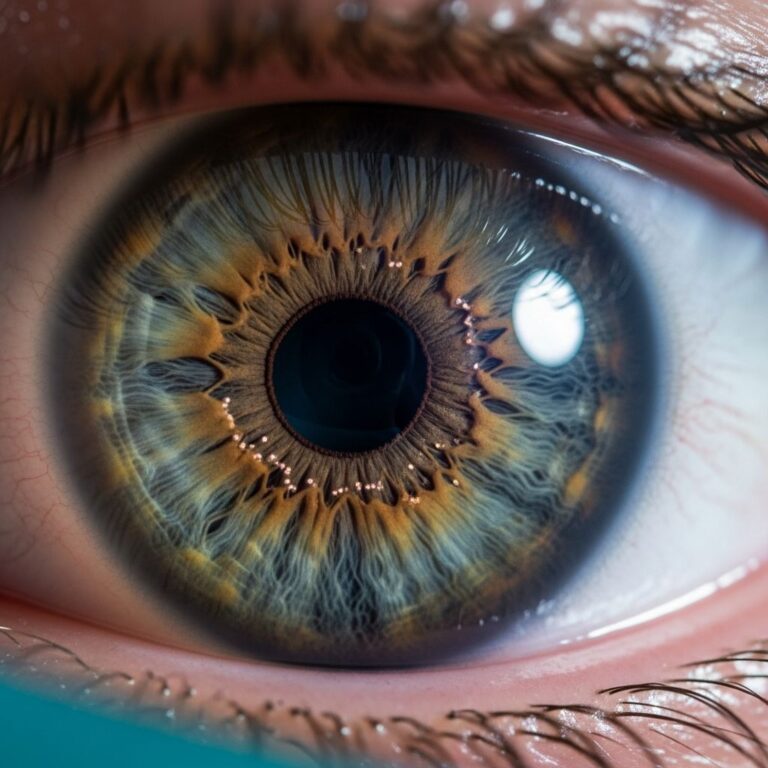
After passing through the cornea, light travels through the aqueous humor, a clear fluid that maintains the eye’s shape and nourishes the cornea and lens. The light then passes through the pupil, the dark circular opening in the center of your iris. The iris, the colored part of your eye, controls how much light enters by expanding and contracting the pupil size. In bright conditions, your pupil constricts to limit light exposure; in dim lighting, it expands to allow more light to reach the retina.
Next, light enters the lens, a transparent structure that fine-tunes the focus of light rays onto the retina. Unlike the cornea, which provides most of your eye’s focusing power, the lens continuously adjusts its shape to maintain sharp focus on objects at varying distances. This process, called accommodation, allows you to see clearly whether you’re reading a book or looking at distant mountains. With age, the lens becomes less flexible, which is why many people develop presbyopia, a condition making it harder to focus on nearby objects.
Light then passes through the vitreous humor, a gel-like substance that fills the majority of your eye’s interior and helps maintain its round shape. Finally, light reaches the retina, a light-sensitive tissue lining the back of your eye. The retina contains millions of specialized cells called photoreceptors—rods and cones—that convert light energy into electrical signals.
The Role of Rods and Cones in Vision
The retina’s photoreceptor cells are divided into two main types: rods and cones, each serving different functions in your vision.
Rods are extremely sensitive to light and are primarily responsible for vision in low-light conditions. They don’t perceive color but are excellent at detecting movement and providing peripheral vision. Rods are concentrated around the edges of the retina, which explains why your peripheral vision is more sensitive to motion than your central vision.
Cones function best in bright light conditions and are responsible for color vision and sharp, detailed sight. They are concentrated in the central retina, particularly in an area called the macula, which provides your sharpest vision. There are three types of cones, each sensitive to different wavelengths of light: red, green, and blue. Your brain combines signals from these three cone types to create your perception of all colors.
When rods and cones detect light, they trigger a chemical reaction that generates electrical signals. These signals travel through several layers of nerve cells in the retina before reaching the optic nerve.
The Optic Nerve and Brain Processing
The optic nerve serves as the communication highway between your eye and brain, transmitting electrical signals generated by photoreceptors to the visual cortex, the part of your brain that processes visual information. Each eye has one optic nerve, and remarkably, the optic nerves from both eyes partially cross at a location called the optic chiasm. This crossing allows your brain to receive information from both eyes and create a unified, three-dimensional image of your world.
Once signals reach the visual cortex in the back of your brain, sophisticated processing occurs. Your brain interprets the signals, combining information from both eyes to assess depth, distance, and the spatial relationships between objects. Your brain also draws on memory and experience to recognize what you’re seeing, making sense of colors, shapes, and movements. This is why vision isn’t purely a mechanical process but involves active interpretation by your brain.
Understanding Visual Acuity
Visual acuity refers to the sharpness and clarity of your vision—your ability to see fine details and distinguish objects from their surroundings. It’s commonly measured on a scale, with 20/20 vision considered standard or normal visual acuity for adults. This measurement means that from a distance of 20 feet, you can see what an average person should be able to see at that distance.
However, visual acuity can vary significantly among individuals. Some people have better than 20/20 vision, such as 20/15 vision, meaning they can see clearly from 20 feet away what requires 15 feet for someone with standard vision. Others may have 20/40 vision, meaning they need to be 20 feet away to see what someone with standard vision can see from 40 feet away.
Several factors influence your visual acuity, including the shape of your cornea and lens, the health of your retina, and the strength of your eye muscles. Refractive errors—conditions where light doesn’t focus properly on the retina—are among the most common causes of reduced visual acuity.
Common Refractive Errors Affecting Vision
Refractive errors occur when the shape of your eye prevents light from focusing directly on the retina, resulting in blurred vision at certain distances.
Myopia (Nearsightedness) occurs when the cornea is too curved or the eyeball is too long, causing light to focus in front of the retina. People with myopia can see nearby objects clearly but have difficulty seeing distant objects. This is one of the most common vision problems worldwide and often develops in childhood.
Hyperopia (Farsightedness) happens when the cornea is too flat or the eyeball is too short, causing light to focus behind the retina. People with hyperopia can typically see distant objects more clearly than nearby objects, though significant hyperopia can blur both near and far vision.
Astigmatism occurs when the cornea or lens has an irregular shape, causing blurred vision at all distances. Rather than being evenly curved, the cornea or lens has different curvatures in different meridians, similar to the shape of a football rather than a sphere.
Presbyopia develops with age as the lens becomes less flexible and loses its ability to change shape for focusing on nearby objects. This typically becomes noticeable around age 40 and affects nearly everyone eventually. Unlike the other refractive errors, presbyopia is a natural part of aging rather than a structural problem.
Other Factors Affecting Vision Quality
Beyond refractive errors, numerous other factors can affect your vision quality and overall eye health.
Aging brings natural changes to the eye, including reduced pupil size, yellowing of the lens, and decreased light sensitivity. These changes are normal but can gradually reduce visual acuity and make night driving more challenging.
Lighting conditions significantly impact vision. Adequate lighting helps your cones function optimally for clear, detailed vision. Poor lighting causes reliance on rods, which provide less detailed vision. Excessive glare can also impair vision by overwhelming the photoreceptors.
Focus and concentration affect how clearly you perceive your environment. When you concentrate on an object, your eye muscles adjust to maintain sharp focus, and your brain processes visual information more thoroughly.
Eye health conditions such as cataracts, glaucoma, macular degeneration, and diabetic retinopathy can significantly impact vision quality. Cataracts cloud the lens, glaucoma damages the optic nerve, and macular degeneration affects the central retina’s function.
Maintaining Healthy Vision
Protecting your vision requires a multifaceted approach to eye health. Regular comprehensive eye exams allow optometrists and ophthalmologists to detect vision problems early and monitor eye health. Wearing appropriate corrective lenses or contact lenses as prescribed ensures optimal visual acuity for daily activities.
Protective eyewear is essential when engaging in activities with eye injury risk, such as sports, home repairs, or workplace tasks. UV protection is equally important—wearing sunglasses that block 100% of UVA and UVB rays prevents cumulative damage from sun exposure.
A healthy diet rich in antioxidants, vitamins, and minerals supports eye health. Nutrients like lutein, zeaxanthin, vitamins C and E, zinc, and omega-3 fatty acids are particularly beneficial for maintaining retinal function and reducing the risk of age-related eye diseases.
Managing systemic conditions like diabetes and high blood pressure is crucial, as these significantly impact vision. Diabetes increases the risk of diabetic retinopathy, while high blood pressure can damage blood vessels in the retina. Regular exercise and maintaining a healthy weight also contribute to overall eye health.
Limiting screen time and practicing the 20-20-20 rule—every 20 minutes, look at something 20 feet away for 20 seconds—reduces eye strain from digital devices. Avoiding smoking is also important, as smoking increases the risk of developing cataracts and age-related macular degeneration.
When to See an Eye Care Professional
Certain symptoms warrant prompt attention from an eye care professional. These include sudden changes in vision, eye pain, persistent redness or irritation, flashing lights or sudden floaters, or vision loss in any portion of your visual field. Regular eye exams are also important for detecting asymptomatic conditions early.
Frequently Asked Questions
Q: What is the difference between 20/20 vision and perfect vision?
A: 20/20 vision is considered standard or normal visual acuity, but it doesn’t account for other aspects of vision like peripheral vision, depth perception, or color vision. Some people have better than 20/20 vision, such as 20/15 vision, which is sometimes called
Similar Articles


Read full bio of medha deb