Von Hippel-Lindau Disease: Causes, Symptoms, Diagnosis & Treatment
Understanding VHL disease: genetic causes, clinical manifestations, surveillance strategies, and modern treatment options.

What is Von Hippel-Lindau Disease?
Von Hippel-Lindau (VHL) disease is a rare hereditary neurocutaneous disorder characterized by the development of benign and malignant tumors across multiple organ systems. This genetic condition predisposes individuals to the formation of blood vessel tumors and various cystic lesions throughout the body, including the eyes, brain, spinal cord, pancreas, kidneys, and other organs. VHL is inherited in an autosomal dominant pattern, meaning that only one copy of the mutated gene is necessary for disease manifestation. Individuals with VHL have a 50% chance of passing the condition to each of their children.
The disease results from mutations in the VHL tumor suppressor gene, located on chromosome 3. This gene normally produces a protein that helps regulate cellular growth and prevents tumor formation. When this gene is defective, the body loses this protective mechanism, leading to uncontrolled cellular proliferation and tumor development across various tissues.
Understanding the Genetic Basis
The VHL gene functions as a tumor suppressor, producing a protein responsible for marking damaged proteins for destruction and regulating oxygen-dependent cellular processes. When mutations occur in this gene, the protective mechanism fails, allowing abnormal cells to multiply unchecked. Different types of VHL mutations result in varying patterns of tumor development, influencing which organs are most likely to be affected and the severity of manifestations.
Mutation variants of VHL disease include Type 1, Type 1B, and Type 2C, each associated with distinct clinical presentations:
| Mutation Type | Genetic Characteristics | High-Risk Manifestations | Low-Risk Manifestations |
|---|---|---|---|
| Type 1 | Deletions, insertions, truncations, missense mutations | CNS hemangioblastoma, retinal hemangioblastoma, renal cell carcinoma, pancreatic neuroendocrine tumor | Pheochromocytoma |
| Type 1B | Gene deletions encompassing VHL | CNS hemangioblastoma, retinal hemangioblastoma, pancreatic neuroendocrine tumors | Renal cell carcinoma, pheochromocytoma |
| Type 2C | Missense mutations (e.g., p.V84L, p.L188V) | Pheochromocytoma | CNS hemangioblastoma, retinal hemangioblastoma, renal cell carcinoma |
Clinical Manifestations and Associated Tumors
VHL disease manifests across multiple organ systems with a wide spectrum of lesions. Clinical symptoms depend on the location and size of affected lesions, with presentation typically occurring between ages 10 and 30, though earlier manifestations are possible. Retinal and central nervous system (CNS) hemangioblastomas generally present earlier in the disease course compared to visceral lesions such as renal cell carcinoma.
Common Manifestations and Lifetime Risk
The most prevalent manifestations of VHL disease include:
| Organ System | Manifestation Type | Lifetime Risk (%) |
|---|---|---|
| Renal System | Renal mass | 25–75 |
| Renal cyst | 42 | |
| Renal cell carcinoma | 17–75 | |
| Pancreas | Pancreatic mass | 35–75 |
| Pancreatic cyst | 75–85 | |
| Pancreatic System | Neuroendocrine tumor | 10–17 |
| Adrenal Glands | Pheochromocytoma and paraganglioma | 10–25 |
| Male Reproductive | Epididymal cystadenoma | 25–60 |
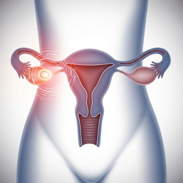
| Female Reproductive | Broad ligament cyst | 10 |
| Inner Ear | Endolymphatic sac tumor | 4–16 |
Retinal Hemangioblastomas
Retinal hemangioblastomas are among the most common manifestations of VHL disease, appearing as dilated arteries leading from the optic disk to peripheral tumors with engorged veins. While typically asymptomatic, centrally located and enlarging lesions can result in substantial vision loss. These tumors increase the risk of retinal detachment, macular edema, and glaucoma. Early detection through regular ophthalmologic examination is crucial to prevent visual deterioration.
Central Nervous System Hemangioblastomas
CNS hemangioblastomas occur most commonly in the cerebellum but can develop throughout the brain and spinal cord. These vascular tumors may cause symptoms including headaches, dizziness, weakness, ataxia, and neurological dysfunction depending on their size and location. Untreated CNS hemangioblastomas can result in brain damage or death.
Renal Manifestations
Renal involvement represents a major concern in VHL disease, with risk of developing renal cell carcinoma increasing significantly with age, potentially reaching 70% by age 60. Patients typically develop multiple renal cysts and masses that require careful monitoring and management to prevent progression to malignancy.
Pancreatic Involvement
Pancreatic manifestations include simple cysts, which are highly prevalent, and neuroendocrine tumors that require surveillance for malignant potential. The high frequency of pancreatic cysts (75-85% lifetime risk) necessitates regular imaging to distinguish benign cystic lesions from tumors requiring intervention.
Pheochromocytomas and Endocrine Abnormalities
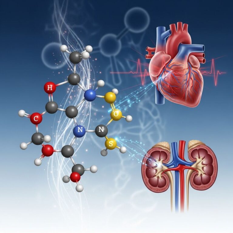
Pheochromocytomas develop in 10-25% of VHL patients, producing excessive catecholamines and leading to hypertension, headaches, and cardiovascular complications. Blood pressure monitoring and biochemical screening are essential for early detection and management.
Diagnosis of Von Hippel-Lindau Disease
Accurate diagnosis of VHL disease hinges on clinical features combined with genetic confirmation of pathogenic mutations in the VHL gene. The diagnostic process involves comprehensive evaluation and multiple imaging modalities.
Diagnostic Criteria
Von Hippel-Lindau disease is diagnosed when one of the following criteria is met:
- Family history of VHL and presence of at least one VHL-associated tumor (retinal, brain, or spinal hemangioblastoma; pheochromocytoma; renal cell carcinoma; or pancreatic endocrine tumor)
- Presence of two or more characteristic VHL tumors with no known family history
- Genetic confirmation of a pathogenic VHL gene mutation
Diagnostic Evaluation Methods
Patients suspected of VHL disease should undergo comprehensive evaluations including:
- Magnetic Resonance Imaging (MRI): MRI with contrast enhancement is the modality of choice for detecting CNS hemangioblastomas along the entire craniospinal axis, including the internal auditory canal
- Ophthalmoscopy: Direct ophthalmoscopy enables visualization and characterization of retinal angiomas and hemangioblastomas
- Abdominal Imaging: Computed tomography (CT) or MRI identifies renal cysts, renal cell carcinomas, pancreatic lesions, and pheochromocytomas
- Biochemical Assessments: Blood pressure monitoring and measurement of urine or plasma metanephrines detect pheochromocytomas
- Genetic Testing: Molecular genetic testing confirms VHL gene mutations, with family history analysis essential for comprehensive assessment
Surveillance and Screening Guidelines
Early detection is crucial to preventing serious complications in VHL disease. The penetrance of VHL disease is nearly complete by age 65 years, after which patients develop few new lesions or symptomatic manifestations. Affected individuals and at-risk relatives should remain under regular surveillance until their seventh decade of life.
Recommended Surveillance Protocol
Routine surveillance for diagnosed VHL patients should include the following:
- Annual History and Physical Examination: Comprehensive clinical assessment for new symptoms and manifestations
- Ophthalmologic Examination: Annual dilated eye examination beginning in infancy to screen for retinal hemangioblastomas, with binocular funduscopy recommended within the first year of life
- Blood Pressure Monitoring: Annual monitoring beginning at age 2 years to screen for pheochromocytomas
- Biochemical Screening: Annual measurement of urine or plasma fractionated metanephrines beginning at age 5 years
- Neuroimaging: Brain and spinal MRI every 2 years beginning at age 11 years to screen for CNS hemangioblastomas
- Audiography: Testing every 2 years beginning at age 11 years to screen for endolymphatic sac tumors
- Abdominal Imaging: MRI or ultrasonography every 2 years beginning at age 15 years to screen for renal cell carcinomas, pheochromocytomas, and pancreatic tumors
For asymptomatic patients over age 30, routine eye examinations can be performed annually, while abdominal imaging recommendations have been adjusted to begin at age 15 with 2-year intervals between studies.
Treatment and Management Approaches
Treatment of VHL disease involves multiple strategies tailored to specific tumor types and individual patient characteristics, focusing on early intervention and prevention of complications.
Surgical Intervention
Surgery remains a cornerstone of VHL management. Treatment often involves surgical removal of tumors before they become harmful. Specific surgical approaches include:
- Pheochromocytoma Surgery: Surgical removal is the primary treatment, with continued management of hypertension required in some patients
- Renal Cell Carcinoma Management: Surgical resection of tumors; advanced cancers may respond to pharmacologic treatment
- CNS Hemangioblastomas: Surgery is performed when tumors become symptomatic or show significant growth
Retinal and Ophthalmic Treatment
For retinal angiomas, several minimally invasive treatments are available depending on lesion size:
- Laser Photocoagulation: Effective for smaller lesions with minimal complications
- Cryotherapy (Freezing Treatment): Alternative approach for smaller lesions to control disease progression
- Intravitreal Injections: Anti-vascular endothelial growth factor medications similar to those used in macular degeneration for larger lesions to control exudation
Management primarily focuses on laser photocoagulation or cryotherapy, which effectively treats vascular lesions with minimal complications. Early detection remains crucial to prevent visual deterioration.
Radiation Therapy
Some tumors can be treated with focused high-dose radiation therapy as an alternative or adjunctive approach when surgery is not feasible or appropriate.
Pharmacologic Treatment: Belzutifan
Recent therapeutic advances have introduced belzutifan, a hypoxia-inducible factor-2α (HIF-2α) inhibitor approved by the Food and Drug Administration for selected VHL patients. This medication represents a significant breakthrough in VHL management, offering systemic treatment for patients with renal cell carcinomas, central nervous system hemangioblastomas, or pancreatic endocrine tumors. This novel approach addresses the underlying molecular mechanisms of VHL disease by targeting hypoxia signaling pathways.
Living with Von Hippel-Lindau Disease
Individuals with VHL disease require comprehensive, multidisciplinary care involving ophthalmologists, neurologists, urologists, endocrinologists, and genetic counselors. Regular surveillance and proactive management significantly improve outcomes and quality of life. Genetic counseling is essential for patients and at-risk family members to understand inheritance patterns, screening recommendations, and reproductive implications.
Untreated VHL can result in blindness, brain damage, or death, with mortality typically resulting from complications of cerebellar hemangioblastomas or renal cell carcinoma. However, with appropriate surveillance and timely intervention, many complications can be prevented or effectively managed.
Frequently Asked Questions
Q: At what age do VHL symptoms typically appear?
A: While VHL manifestations typically appear between ages 10 and 30, symptoms can develop earlier. Retinal and CNS hemangioblastomas generally present before visceral lesions like renal cell carcinoma. Regular screening beginning in infancy is recommended for diagnosed or at-risk individuals.
Q: How is VHL disease inherited?
A: VHL disease follows an autosomal dominant inheritance pattern, meaning only one mutated copy of the VHL gene is necessary for disease development. Individuals with VHL have a 50% chance of passing the condition to each child. Approximately 20% of cases result from new mutations without family history.
Q: Can VHL disease be cured?
A: Currently, there is no cure for VHL disease, as it is a genetic condition affecting all cells in the body. However, regular surveillance, early detection, and timely treatment of tumors can significantly improve outcomes and prevent serious complications. New therapeutic options like belzutifan offer improved management strategies.
Q: What is the life expectancy for someone with VHL disease?
A: Life expectancy varies based on the type of VHL mutation, surveillance adherence, and timely treatment. With comprehensive surveillance programs and modern treatment options, many VHL patients achieve normal or near-normal life expectancy. However, untreated complications from CNS hemangioblastomas or renal cell carcinoma can be fatal.
Q: How often should VHL patients be screened?
A: Screening frequency depends on age and manifestation type. Generally, annual comprehensive examinations are recommended, with specific imaging intervals (ophthalmology annually, MRI every 2 years starting at age 11, abdominal imaging every 2 years starting at age 15). Surveillance intensity may decrease after age 65 when disease penetrance is complete.
Q: What should at-risk family members of VHL patients do?
A: At-risk relatives should undergo genetic testing and, if positive, follow the same comprehensive surveillance protocol as diagnosed patients. Even untested first- and second-degree relatives should be screened, beginning in childhood, as early detection of tumors significantly improves outcomes.
References
- Von Hippel-Lindau Disease: A Comprehensive Review — National Center for Biotechnology Information (NCBI). 2024. https://pmc.ncbi.nlm.nih.gov/articles/PMC12062539/
- Von Hippel-Lindau Disease (VHL) – Pediatrics — Merck Manuals Professional Edition. 2025. https://www.merckmanuals.com/professional/pediatrics/neurocutaneous-syndromes/von-hippel-lindau-disease-vhl
- Von Hippel-Lindau (VHL) Disease: Blood Vessel Tumors — Johns Hopkins Medicine. 2024. Available through institutional educational resources.
Similar Articles


Read full bio of medha deb