Wells Syndrome Pathology: Understanding Eosinophilic Infiltration
Comprehensive guide to Wells syndrome pathology, histopathological findings, and diagnostic criteria for healthcare professionals.

Wells Syndrome Pathology: Understanding Eosinophilic Infiltration and Histological Features
nn
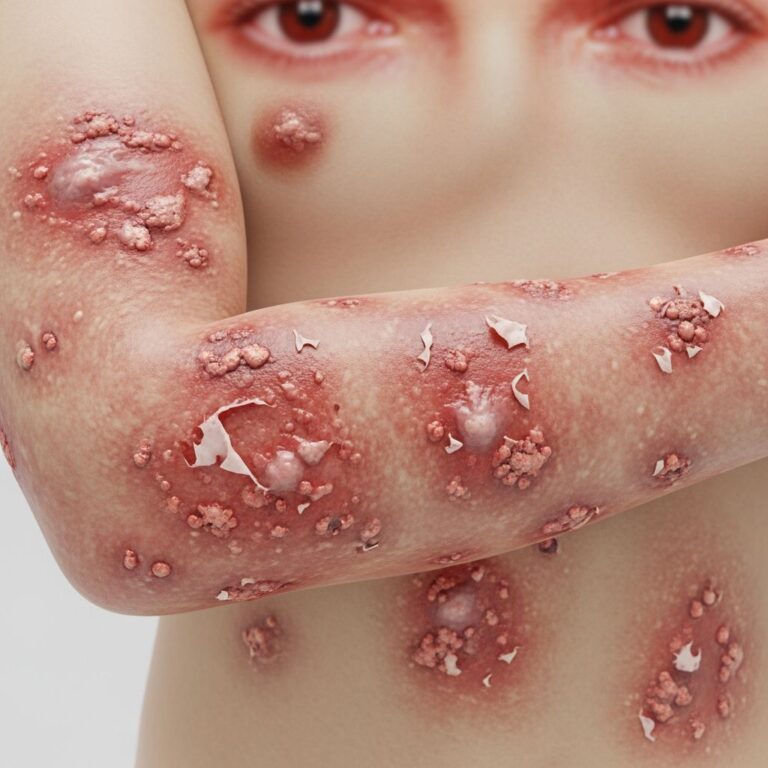
Wells syndrome, clinically known as eosinophilic cellulitis, is an uncommon inflammatory skin condition characterized by distinctive pathological features that differentiate it from infectious cellulitis. Understanding the pathology of Wells syndrome is crucial for dermatologists and healthcare professionals to establish accurate diagnosis and implement appropriate treatment strategies. The condition was first identified by Dr. George C. Wells in 1971 as a recurrent granulomatous dermatitis with eosinophilia, and since then, fewer than 200 cases have been documented in medical literature.
nn
Histopathological Features and Microscopic Findings
nn
The histopathological examination of Wells syndrome reveals characteristic and distinctive microscopic findings that form the gold standard for diagnosis. Dermal edema and eosinophilic infiltration are present in virtually all cases, occurring in 100% of documented patients. These findings, combined with additional pathological markers, create a recognizable pattern that distinguishes Wells syndrome from other inflammatory dermatoses.
nn
The classic histopathological presentation includes the following key features:
nn
- n
- Dense eosinophilic infiltration in the dermis, extending into the subcutaneous tissue
- Perivascular eosinophilic infiltrate surrounding blood vessels
- Interstitial dermal infiltration with eosinophils distributed throughout dermis layers
- Dermal edema contributing to tissue swelling and inflammation
- Characteristic “flame figures” present in the majority of cases (96%)
n
n
n
n
n
nn
Understanding Flame Figures: The Pathological Hallmark
nn
Flame figures represent the most distinctive histopathological feature of Wells syndrome and serve as a crucial diagnostic marker. These structures consist of collagen fibrils coated with eosinophilic granule proteins, creating a characteristic appearance under microscopic examination. The flame figures result from the degranulation of eosinophils within the dermis, where granular material from eosinophils adheres to collagen bundles and surrounding tissue.
nn
The composition and formation of flame figures involve:
nn
- n
- Eosinophil major basic protein identified within granules through immunofluorescent staining
- Amorphous or granular eosinophilic material coating collagen fibers
- Phagocytic histiocytes engulfing eosinophilic material
- Formation occurring during the subacute phase of inflammation
n
n
n
n
nn
While flame figures are characteristic of Wells syndrome, it is important to recognize that they are not pathognomonic. These structures can also be observed in other eosinophil-rich infiltrate conditions, including insect bite reactions, bullous pemphigoid, and Churg-Strauss syndrome. Therefore, flame figures must be interpreted within the context of clinical presentation and other histopathological findings.
nn
Pathological Stages of Disease Development
nn
Wells syndrome progresses through distinct histopathological stages, each characterized by specific microscopic features and inflammatory patterns. Understanding these phases provides insight into the disease mechanism and helps correlate clinical presentation with underlying pathology.
nn
Early Phase
nn
The early phase of Wells syndrome is characterized by prominent dermal edema and widespread distribution of eosinophils throughout the dermis. During this initial stage, inflammation appears acute with eosinophilic cells dominating the infiltrate. The lesions typically present with erythema, edema, and clinical features resembling infectious cellulitis, which frequently leads to initial misdiagnosis.
nn
Subacute Phase
nn
The subacute phase represents the transition period where characteristic flame figures become prominent. During this stage, phagocytic histiocytes appear and begin engulfing eosinophilic material. The infiltrate becomes more organized, with amorphous or granular eosinophilic material distinctly adhering to collagen bundles. This phase typically coincides with clinical induration of plaques and the development of well-defined borders.
nn
Chronic Phase
nn
In the chronic phase, eosinophils become fewer in number, and the histopathological pattern shifts toward a more granulomatous appearance. Giant cells may be observed between collagen bundles alongside remaining histiocytes. The flame figures may begin to disappear, and granulomatous infiltrate becomes more prominent. This phase correlates with clinical resolution and the beginning stages of wound healing, though recurrence is common.
nn
Pathophysiology and Immunological Mechanisms
nn
The underlying mechanism of Wells syndrome involves dysregulated eosinophilia driven by abnormal immune responses. Research into the immunopathology reveals important insights into disease initiation and progression.
nn
Key immunological features include:
nn
- n
- T-cell involvement: Elevated percentages of CD3+ and CD4+ T-cells in peripheral blood and tissue
- Cytokine release: Spontaneous release of Interleukin 5 (IL-5) in substantial quantities by T-cells, a critical factor in eosinophil recruitment and activation
- Eosinophil degranulation: Activated eosinophils release granule contents containing major basic protein, causing tissue damage and inflammation
- Edema formation: Released granule proteins trigger increased vascular permeability and fluid accumulation in tissues
n
n
n
n
nn
Interleukin 5 plays a central role in the pathophysiology of Wells syndrome, as it is the primary cytokine involved in promoting both blood and tissue eosinophilia. The dysregulation of this IL-5-mediated eosinophil response appears fundamental to disease development, though the trigger initiating this abnormal response remains incompletely understood.
nn
Cellular Infiltrate Composition and Organization
nn
The dermal infiltrate in Wells syndrome comprises multiple cell types organized in characteristic patterns. The composition varies depending on the disease stage but consistently includes specific cellular elements that contribute to the distinctive histopathological picture.
nn
| Cell Type | Location in Dermis | Function | Temporal Pattern |
|---|---|---|---|
| Eosinophils | Diffuse dermal and perivascular | Primary inflammatory cells; release granule proteins | Prominent in early and subacute phases; decrease in chronic phase |
| Histiocytes (macrophages) | Interstitial dermis and between collagen bundles | Phagocytose eosinophilic material; form granulomatous response | Increase during subacute and chronic phases |
| Giant cells | Between collagen bundles | Part of granulomatous response; process foreign material | Appear in chronic phase |
| T-lymphocytes | Perivascular and interstitial | Drive eosinophil recruitment through cytokine secretion | Present throughout disease course |
nn
Differential Pathological Diagnosis
nn
Several dermatological conditions can present with eosinophilic infiltrates, making differential diagnosis essential for accurate identification of Wells syndrome. While flame figures are characteristic of Wells syndrome, they are not exclusive, and several conditions must be considered when evaluating eosinophil-rich infiltrates.
nn
Conditions presenting similar histopathological features include:
nn
- n
- Insect bite reactions: Show eosinophilic infiltrate with possible flame figures but differ in clinical presentation and distribution
- Bullous pemphigoid: May display eosinophil-rich infiltrates and occasional flame figures but present with distinct subepidermal blistering
- Churg-Strauss syndrome: Systemic vasculitis with eosinophil infiltration and necrotizing vasculitis, distinguished by systemic manifestations and vascular involvement
- Eosinophilic granuloma: Predominantly histiocytic infiltrate with variable eosinophilic component
- Parasitic infections: Eosinophil-rich infiltrates related to parasitic organisms, identifiable through specific staining
n
n
n
n
n
nn
Clinical-Pathological Correlation
nn
The relationship between histopathological findings and clinical presentation in Wells syndrome provides valuable diagnostic information. Early clinical lesions presenting with erythema and edema correspond to the early histopathological phase with prominent dermal edema and eosinophilic infiltration. As lesions progress to indurated plaques with well-defined borders, histopathology shows the subacute phase with characteristic flame figures and histiocytic involvement.
nn
The variable morphology of clinical lesions reflects the heterogeneity of pathological changes. Some patients present with predominantly cellulitis-like plaques, while others develop bullous, nodular, or granuloma-like lesions. This clinical variability correlates with differences in the depth and intensity of eosinophilic infiltration and the degree of dermal edema. Lesions that resolve spontaneously show histopathological evidence of decreased inflammation and transition toward normal tissue architecture, though the potential for recurrence remains due to the underlying dysregulated immune response.
nn
Histopathological Examination Techniques
nn
Proper tissue examination and staining techniques are essential for identifying pathological features of Wells syndrome. Skin biopsy remains the gold standard for diagnosis, with histopathological findings forming the cornerstone of diagnostic confirmation.
nn
Recommended examination approaches include:
nn
- n
- Hematoxylin and eosin (H&E) staining: Standard staining reveals eosinophils, dermal edema, and overall architectural changes
- Immunofluorescent staining: Identifies eosinophil major basic protein within flame figures and granule material
- Immunohistochemical stains: May identify specific T-cell subsets (CD3, CD4) and other inflammatory markers
- Multiple tissue sections: Examination of serial sections increases likelihood of identifying flame figures and representative pathological changes
n
n
n
n
nn
Evolution of Histopathology Over Time
nn
As individual lesions of Wells syndrome progress through their natural course, histopathological changes continue to evolve. Early lesions show acute inflammation, whereas older lesions display evidence of tissue remodeling and reduced inflammatory burden. This temporal evolution has important implications for biopsy timing and interpretation. Biopsies obtained from newer lesions are more likely to demonstrate characteristic early-phase findings with prominent eosinophilic infiltration, while specimens from resolving lesions may show predominantly granulomatous features with less obvious flame figures.
nn
The persistent potential for disease recurrence, despite clinical resolution and apparent histopathological healing, suggests that complete immunological normalization may not occur. Residual abnormalities in eosinophil regulation or T-cell dysfunction likely persist even after clinical clearance, predisposing affected individuals to future flare-ups.
nn
Frequently Asked Questions
nn
Q: Are flame figures pathognomonic for Wells syndrome?
nn
A: No, flame figures are characteristic but not pathognomonic. While present in 96% of Wells syndrome cases, they can also occur in other eosinophil-rich conditions including insect bite reactions, bullous pemphigoid, and Churg-Strauss syndrome. Diagnosis requires correlation with clinical presentation and other histopathological features.
nn
Q: How is Wells syndrome histopathology distinguished from infectious cellulitis?
nn
A: Infectious cellulitis shows bacterial infiltration with neutrophilic rather than eosinophilic inflammation, lacks flame figures, and typically presents as acute infection. Wells syndrome demonstrates eosinophil-predominant infiltrate with characteristic flame figures and lacks identifiable infectious organisms. Culture and gram staining of Wells syndrome specimens are negative for bacteria.
nn
Q: What role do T-cells play in Wells syndrome pathology?
nn
A: T-cells, particularly CD4+ and CD3+ subsets, drive the immunological abnormality by spontaneously releasing IL-5. This cytokine recruits and activates eosinophils, which subsequently degranulate and damage tissue. The dysregulation of this T-cell driven response appears fundamental to disease pathogenesis.
nn
Q: Why does Wells syndrome recur despite apparent histopathological healing?
nn
A: Although individual lesions heal with resolution of acute inflammation and reduction of eosinophilic infiltrate, the underlying T-cell dysregulation and abnormal IL-5 production persist. This allows for recurrent episodes of eosinophil recruitment and activation, explaining the characteristic relapsing-remitting course of the disease.
nn
Q: How does biopsy timing affect histopathological findings in Wells syndrome?
nn
A: Biopsies from newer lesions are more likely to show early-phase findings with prominent eosinophilic infiltration and dermal edema. Older, resolving lesions demonstrate subacute and chronic phase changes with more prominent granulomatous features and potentially fewer visible flame figures. For diagnostic purposes, biopsy of active lesions is recommended.
nn
References
n
- n
- An Itchy Mystery: A Rare Case of Wells Syndrome — Cosmoderma. 2024. https://cosmoderma.org/an-itchy-mystery-a-rare-case-of-wells-syndrome/
- Diagnosis and Management of Eosinophilic Cellulitis (Wells’ Syndrome) — National Center for Biotechnology Information (NCBI/PMC). 2012. https://pmc.ncbi.nlm.nih.gov/articles/PMC3383552/
- Wells Syndrome (Eosinophilic Cellulitis) — DermNet New Zealand. 2024. https://dermnetnz.org/topics/wells-syndrome
n
n
n
n
n
Similar Articles


Read full bio of Sneha Tete