What Does Squamous Cell Carcinoma Look Like?
Recognize squamous cell carcinoma early: Identify scaly lumps, open sores, and other key signs of this common skin cancer for timely treatment.

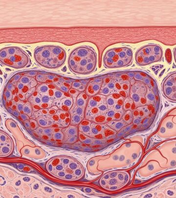
Squamous cell carcinoma (SCC), the second most common form of skin cancer after basal cell carcinoma, typically presents as a gradually enlarging, scaly or crusty lump on the skin. These lesions often develop on sun-exposed areas such as the face, ears, neck, lips, scalp, hands, and forearms, though they can arise anywhere, including scars or chronic wounds. Early detection is critical, as SCC is highly curable when treated promptly but can invade deeper tissues or metastasize if neglected.
SCC arises from malignant transformation of keratinocytes, the predominant cells in the epidermis. Risk factors include cumulative ultraviolet (UV) radiation exposure, fair skin, immunosuppression, chronic wounds, and human papillomavirus (HPV) infection. Lesions grow over weeks to months, varying from millimeters to several centimeters in diameter. Unlike benign conditions, SCC lesions persist, evolve, and may bleed, ulcerate, or cause pain.
Clinical features of squamous cell carcinoma
SCC exhibits diverse morphologies, often mimicking benign conditions like actinic keratosis, warts, or eczema, which underscores the need for professional evaluation. Common features include:
- Scaly or crusted lumps: Firm, hyperkeratotic nodules with a rough, adherent scale that may flake off to reveal a tender base.
- Open sores (ulcers): Non-healing erosions or ulcers with rolled edges, often oozing, crusted, or bleeding intermittently.
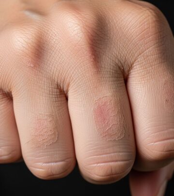
- Red or inflamed patches: Persistent, rough, scaly plaques that may itch, bleed, or develop a central depression.
- Wart-like growths: Verrucous, exophytic proliferations resembling common warts but with rapid growth and asymmetry.
- Dome-shaped or nodular growths: Raised, firm papules or nodules, sometimes with a central keratin plug or crust.
These features align with descriptions from authoritative sources: the American Cancer Society notes rough, scaly red patches, raised growths, and recurrent sores as hallmarks. Similarly, the Skin Cancer Foundation emphasizes thick, scaly patches that crust or bleed. In darker skin types, SCC may present as darker-hued lesions but retains similar textural changes.
Colours of squamous cell carcinoma
SCC lesions display a spectrum of colors influenced by pigmentation, inflammation, and keratinization:
- Red or pink: Most common due to vascularity and inflammation, especially in early or superficial lesions.
- Brown, tan, or black: From melanin in pigmented skin or deeper invasion.
- Pearly white or yellow: Hyperkeratotic areas resembling horns or cutaneous horns.
- Grey or variegated: In advanced or ulcerated tumors with necrosis.
According to DermNet, SCCs can be a range of different colours. The American Academy of Dermatology highlights reddish-brown scaly bumps, while MD Anderson notes tender, raised areas that bleed easily. Color variability complicates self-diagnosis, as benign seborrheic keratoses or seborrheic dermatitis can appear similar.
Size of squamous cell carcinoma
SCC size at presentation varies widely:
- Early lesions: 3–10 mm, often subtle and mistaken for actinic damage.
- Established tumors: 1–5 cm, with potential for further expansion.
- Neglected cases: Over 5 cm, associated with higher metastasis risk.
Growth is typically rapid, progressing over weeks to months, distinguishing SCC from slower-growing basal cell carcinomas. University of Michigan Health describes firm bumps or scaly patches that enlarge steadily.
How quickly does squamous cell carcinoma grow?
SCC demonstrates aggressive local growth:
- Typical timeline: Noticeable enlargement within 1–3 months.
- High-risk variants: Faster progression, invading cartilage or bone within weeks.
- Invasive potential: 2–5% metastasize to lymph nodes or distant sites if untreated.
Baptist Health emphasizes that changes lasting over two weeks warrant evaluation. Frieder Dermatology notes persistent patches that bleed or crust, urging early intervention.
Variations in appearance of squamous cell carcinoma
SCC morphology is pleomorphic, with subtypes including:
| Subtype | Description | Common Sites |
|---|---|---|
| Classic SCC | Scaly nodule or plaque with ulceration | Sun-exposed skin (face, hands) |
| Cutaneous horn | Conical projection of keratin over a persistent base | Scalp, face |
| In situ SCC (Bowen’s disease) | Sharply demarcated red plaque, slow-growing | Lower legs, trunk |
| Verrucous carcinoma | Wart-like, exophytic, locally invasive | Soles, oral mucosa |
| Ulcerative SCC | Deep crateriform ulcer with heaped-up edges | Lips, ears |
These variations are corroborated across sources: The Derm Group describes hard red growths, damaged sunburn-like skin, and sores in atypical locations like genitals. AAD notes dome-shaped growths or non-healing sores.
Squamous cell carcinoma on the lip
Lip SCC, often on the lower lip, appears as:
- Crusted, indurated plaque or ulcer
- White, thickened patch (leukoplakia)
- Raised nodule with central erosion
High metastasis risk due to vascularity; 90%+ cure rate with early excision.
Squamous cell carcinoma in the mouth
Oral SCC presents as:
- Persistent white/red patches (erythroplakia)
- Ulcerated swellings
- Indurated masses on tongue, floor of mouth
Risk factors: tobacco, alcohol, HPV.
Frequently asked questions
What does the beginning of squamous cell carcinoma look like?
Early SCC resembles actinic keratosis: a rough, scaly red patch or subtle firm papule that persists and grows.
Can squamous cell carcinoma be flat?
Yes, in situ forms like Bowen’s disease appear as flat, erythematous plaques.
How can you tell if a spot is squamous cell carcinoma?
Key signs: persistence >2 weeks, growth, bleeding, crusting. Biopsy confirms.
Is squamous cell carcinoma itchy?
Often yes, especially raised patches; pain or tenderness signals deeper invasion.
References
- Squamous Cell Carcinoma | Skin Cancer | Medical Dermatologist — Frieder Dermatology. 2024. https://friederdermatology.com/squamous-cell-carcinoma/
- Basal and Squamous Cell Skin Cancer Symptoms — American Cancer Society. 2025-01-15. https://www.cancer.org/cancer/types/basal-and-squamous-cell-skin-cancer/detection-diagnosis-staging/signs-and-symptoms.html
- Squamous Cell Carcinoma of the Skin Symptoms & Diagnosis — Baptist Health. 2024. https://baptisthealth.net/services/cancer-care/miami-cancer-institute/our-approach/adult-cancers/skin-cancers/squamous-cell-carcinoma-of-the-skin/symptoms-and-diagnosis
- Skin Cancer: Do You Know the Warning Signs of Squamous Cell Carcinoma? — The Derm Group Partners. 2024. https://thedermgrouppartners.com/skin-cancer-do-you-know-the-warning-signs-of-squamous-cell-carcinoma/
- Squamous Cell Carcinoma Warning Signs and Images — Skin Cancer Foundation. 2025-01-10. https://www.skincancer.org/skin-cancer-information/squamous-cell-carcinoma/scc-warning-signs-and-images/
- What does squamous cell carcinoma look like? — DermNet NZ. 2024-06-01. https://dermnetnz.org/topics/cutaneous-squamous-cell-carcinoma/what-does-squamous-cell-carcinoma-look-like
- Squamous cell carcinoma: From symptoms to treatments — American Academy of Dermatology. 2024. https://www.aad.org/skin-conditions/dermatology-a-to-z/squamous-cell-carcinoma
Similar Articles
Read full bio of medha deb


