White Sponge Naevus: Key Insights On Diagnosis & Management
Rare genetic disorder causing benign white spongy lesions on oral and other mucous membranes, typically asymptomatic and requiring no treatment.

White sponge naevus (WSN), also known as white sponge nevus, is a rare autosomal dominant genetic disorder characterised by the development of benign, white, spongy plaques on mucous membranes, primarily affecting the oral cavity. This condition arises from mutations in the keratin genes KRT4 or KRT13, leading to defective keratin filaments that cause epithelial cell vacuolization and the characteristic spongy appearance.
What is white sponge naevus?
White sponge naevus is an uncommon ectodermal dysplasia primarily manifesting in non-cornifying stratified squamous epithelia such as the buccal mucosa. First described in 1909 by Hyde and named in 1935 by Cannon, it was previously termed congenital leukokeratosis mucosae oris, hereditary leukokeratosis oris, and familial white folded gingivostomatitis. The Mendelian Inheritance in Man (MIM) number is 193900.
It exhibits high penetrance but variable expressivity, meaning affected individuals inherit the mutation but the severity and extent of lesions differ. Lesions result from abnormal keratinisation where suprabasal keratinocytes accumulate keratin intermediate filaments, creating a vacuolated, spongy texture.
Prevalence is unknown due to underreporting, as many cases are asymptomatic and undiagnosed. Some studies suggest a female predominance, though evidence is inconsistent.
Who gets white sponge naevus?
White sponge naevus follows an autosomal dominant inheritance pattern, so it affects males and females equally in theory, with a 50% chance of transmission to offspring. Family history is often positive, aiding clinical suspicion.
- Lesions may be present at birth (neonatal onset) or appear in early childhood, adolescence, or rarely adulthood.
- Penetrance is nearly complete, but expressivity varies widely—from subtle localised patches to extensive mucosal involvement.
- No racial or geographic predisposition is reported.
- Extraoral sites are less common but can involve nasal, oesophageal, laryngeal, genital (vulva, vagina), or anal mucosae.
What causes white sponge naevus?
The condition is caused by heterozygous missense mutations in the keratin cluster genes on chromosome 12q13: specifically KRT4 (keratin 4) or KRT13 (keratin 13). These intermediate filament proteins are expressed in differentiating suprabasal cells of cornifying and non-cornifying epithelia.
Mutations disrupt keratin filament assembly, leading to aggregation, cytoplasmic vacuolisation, and nuclear condensation in keratinocytes. This structural defect produces the macroscopic white, folded, spongy plaques without inflammation or malignant potential.
No environmental triggers are implicated; it is purely genetic.
What are the clinical features of white sponge naevus?
Clinically, white sponge naevus presents as bilateral, symmetrical, soft, white-grey, raised plaques with a folded, corrugated, or velvety surface on mucous membranes. The lesions are thick (2–3 mm), spongy to palpation, rough-textured, and non-scrapable, distinguishing them from pseudomembranous conditions.
Oral manifestations
- Buccal mucosa: Most common site (up to 90% of cases), bilateral from commissures to retromolar pads.
- Other intraoral sites: Labial mucosa, ventral tongue, floor of mouth, soft palate, alveolar ridges (rarely).
- Spared areas: Gingival margins, dorsal tongue, hard palate—key for differential diagnosis.
Extraoral manifestations
- Nasal mucosa, oesophagus, larynx (endoscopy may reveal).
- Genital: Vulvovaginal leukoplakia-like plaques in females; penile or scrotal in males (rare).
- Anal/rectal mucosa.
Lesions are usually asymptomatic but may cause aesthetic concerns or secondary irritation (roughness, burning from superinfection). No skin, nail, hair, or tooth involvement occurs.
Diagnosis of white sponge naevus
Diagnosis combines clinical features, family history, and histopathology; genetic testing confirms but is rarely needed.
Clinical diagnosis
Suspect WSN in bilateral buccal white plaques with spongy texture, positive family history, and no skin/systemic disease.
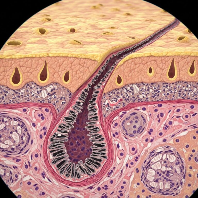
Histopathology
Biopsy is characteristic and confirmatory:
- Epithelial changes: Hyperparakeratosis, acanthosis (thickened spinous layer).
- Key feature: Vacuolised suprabasal keratinocytes with perinuclear eosinophilic condensation (keratin filaments).
- Other: Compact keratin aggregates in upper spinous layers; minimal submucosal inflammation.
Immunohistochemistry shows KRT4/KRT13 expression limited to lesional epithelium.
Differential diagnosis
| Condition | Key Distinguishing Features |
|---|---|
| Leukoplakia | Scrapable? No; risk of dysplasia; tobacco-associated; unilateral often. |
| Candidiasis | Scrapable white plaques; responds to antifungals; erythematous base. |
| Lichen planus | Wickham striae, symmetric, painful, skin involvement possible. |
| Frictional keratosis | History of trauma/cheek biting; resolves with removal of irritant. |
| Hereditary benign intraepithelial dyskeratosis | Bulbar conjunctiva involved; bulbar superior. |
| Pachyonychia congenita |
How is white sponge naevus treated?
White sponge naevus requires no treatment as it is benign, non-progressive, and non-malignant. No reports of malignant transformation exist.
- Symptomatic relief: For secondary bacterial/fungal overgrowth causing burning (stomatopyrosis), use topical antifungals (e.g., clotrimazole) or antibiotics.
- Experimental: Oral doxycycline (anti-inflammatory/keratin-stabilising) showed success in case reports.
- Avoid: Surgery/laser—lesions recur.
Reassurance and regular dental/oral medicine follow-up suffice.
What is the outcome for white sponge naevus?
Lifelong persistence with stable or fluctuating extent; no spontaneous resolution but no complications beyond occasional discomfort. Psychological impact from appearance may prompt reassurance. Genetic counselling for families recommended.
Frequently asked questions (FAQs)
Is white sponge naevus cancerous?
No, it is entirely benign with no malignant potential reported.
Can white sponge naevus be cured?
No cure exists; it is genetic and persistent, but harmless.
Does white sponge naevus affect children?
Yes, often presents from birth or childhood; monitor for secondary infection.
Is biopsy always needed for diagnosis?
Not always if classic clinical/family history, but recommended to exclude mimics.
Can white sponge naevus spread to skin?
No, strictly mucosal; skin is spared.
References
- White sponge nevus — Wikipedia. 2023-10-15. https://en.wikipedia.org/wiki/White_sponge_nevus
- White sponge naevus — DermNet NZ. 2024-01-01. https://dermnetnz.org/topics/white-sponge-naevus
- White sponge naevus: an uncommon oral white lesion — PMC / NIH. 2021-11-23. https://pmc.ncbi.nlm.nih.gov/articles/PMC8634252/
- White Sponge Nevus — European Association of Oral Medicine. 2023-05-12. https://eaom.eu/education/eaom-handbook/white-sponge-nevus/
- White sponge nevus — Orphanet. 2024-02-20. https://www.orpha.net/en/disease/detail/171723
- Successful Treatment of White Sponge Nevus with Oral Doxycycline — Actas Dermo-Sifiliográficas. 2021-04-15. https://www.actasdermo.org/es-successful-treatment-white-sponge-nevus-articulo-S157821902100086X
Similar Articles


Read full bio of medha deb