X-Ray: What It Is, What It Shows, Preparation & Types
Complete guide to X-rays: Understanding how this essential imaging technology works and what it reveals.

What Is an X-Ray?
An X-ray is a type of medical imaging that uses radiation to take pictures of the inside of your body. Often called plain radiographs, X-rays are one of the most common diagnostic tools used in healthcare today. They work by passing a focused beam of radiation through your body, creating black-and-white images that healthcare providers can analyze to diagnose various conditions.
The technology behind X-rays relies on the principle that different tissues in your body absorb radiation at different rates. When X-ray radiation passes through your body, dense structures like bones absorb more radiation and appear white on the image, while soft tissues like muscles and organs allow more radiation to pass through and appear in shades of gray. Air-filled spaces, such as your lungs, allow even more radiation to pass through and appear black on the image.
X-rays are quick, noninvasive tests that provide immediate visual information about what’s happening inside your body. Healthcare providers can use them to make rapid diagnoses, helping you understand whether something concerning needs treatment or additional testing.
What Does an X-Ray Show?
X-rays can reveal a wide range of conditions affecting different parts of your body. Your provider may order an X-ray to investigate various health concerns and monitor existing conditions. Here are some of the main issues that can show up on X-rays:
- Broken bones and fractures
- Arthritis and joint problems
- Infections in your lungs, such as pneumonia
- Spine conditions and abnormalities
- Teeth and dental issues
- Foreign objects that may have been swallowed or lodged in your body
- Heart and lung conditions
- Certain signs of cancer
It’s important to understand that while X-rays are excellent for visualizing bones and some soft tissue conditions, they have limitations. Certain abnormalities don’t always show up on X-rays, even if they’re present. For example, kidney stones and tumors may not be visible on an X-ray image, and cancer detection through X-rays depends on the size and location of the tumor. Small tumors or those hidden behind other structures in your body may not appear on standard X-ray images.
When Might Your Provider Order an X-Ray?
Your healthcare provider may recommend an X-ray for several reasons, depending on your symptoms and medical history. Common scenarios where X-rays are ordered include:
- Suspected broken bones or fractures from injuries
- Evaluation of joint pain or swelling
- Investigation of back or spine pain
- Assessment of respiratory symptoms like cough or difficulty breathing
- Monitoring of chronic conditions like arthritis or COPD
- Evaluation of dental problems
- Post-surgical imaging to ensure proper healing
- Emergency assessment of trauma or injuries
How X-Rays Work: The Technology
Understanding how X-rays work can help ease any concerns you might have about the procedure. X-ray machines produce a focused beam of radiation that passes through your body. The amount of radiation exposure is carefully controlled and minimized to the level necessary to capture quality diagnostic images.
The fundamental principle behind X-ray imaging involves the varying density of body tissues. Your body’s tissues vary significantly in thickness and composition. When radiation passes through your body, each structure allows a different amount of radiation to pass through, creating the characteristic contrast seen in X-ray images.
Dense structures like bones are very thick and don’t allow much radiation to pass through, which is why they appear white on X-ray images. Your lungs, however, contain mostly air and allow much more radiation through, causing them to appear gray. This contrast between different tissue densities allows radiologists to identify abnormalities and structures within your body.
Preparation for an X-Ray
X-rays require minimal preparation compared to many other diagnostic procedures. In most cases, you can proceed with your daily activities as normal and arrive at your appointment ready for the procedure. However, there are a few important considerations:
- Inform your healthcare provider if you are pregnant or might be pregnant
- Remove metal objects, jewelry, and clothing with metal fasteners before the X-ray
- Wear comfortable, easy-to-remove clothing
- Inform the technologist of any recent injuries or relevant medical history
- Stay still during the X-ray to ensure clear images
The procedure itself is quick, usually taking only a few minutes. A trained X-ray technologist will position you appropriately for the area being imaged and may place shielding over sensitive areas like your reproductive organs to minimize radiation exposure.
Common Types of X-Rays
Several types of X-rays take pictures of different areas inside your body. Healthcare providers select the appropriate type based on the area of concern and the diagnostic information needed.
Chest X-Ray (CXR)
A chest X-ray uses a focused beam of radiation to create images of your heart, lungs, and ribcage. This is one of the most commonly ordered X-rays and is often the first test used to diagnose lung and heart conditions. Chest X-rays can help diagnose pneumonia, COPD, emphysema, and other respiratory conditions.
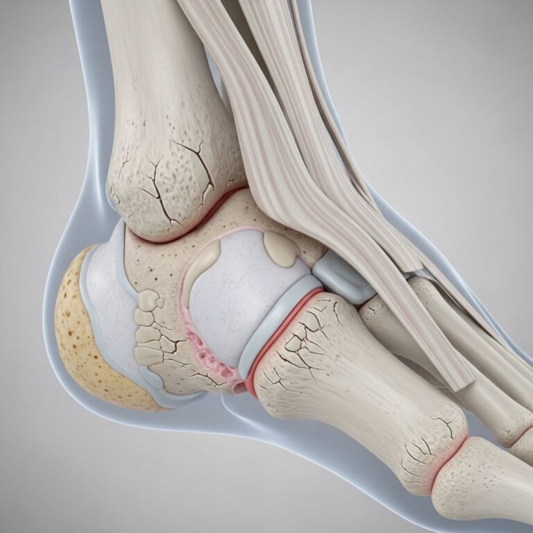
Skeletal X-Rays
These X-rays focus on bones and joints throughout your body. They’re particularly useful for diagnosing fractures, evaluating joint problems, and assessing bone density. Skeletal X-rays can target specific areas like the hand, arm, leg, foot, spine, or pelvis.
Dental X-Rays
Dental X-rays are specialized images used by dentists and dental specialists to examine your teeth and jaw. These small, focused X-rays help identify cavities, infections, and other dental problems.
Abdominal X-Rays
These X-rays image your stomach, intestines, and other abdominal organs. They can help diagnose conditions like bowel obstruction, appendicitis, or other abdominal concerns.
Understanding Your X-Ray Results
After your X-ray is completed, the technologist will send your images to a radiologist—a medical doctor specially trained in interpreting medical images. The radiologist reviews the images for normal and abnormal findings and prepares a detailed report.
Your healthcare provider will then review both the images and the radiologist’s report to discuss your X-ray results with you. In nonemergency cases, you’ll typically receive your results within one to two days. In emergency situations, you’ll usually know your results in a few minutes or hours.
Your provider will explain what the results mean in terms you can understand and discuss any next steps, whether that’s treatment, additional testing, or monitoring. If abnormal results are found, your provider may recommend additional imaging tests like a CT scan or PET scan for more detailed information.
Radiation Safety and X-Rays
One common concern patients have about X-rays involves radiation exposure. It’s important to know that X-rays use a very small amount of radiation—only the necessary amount needed to capture quality diagnostic images. Healthcare providers carefully balance the need for diagnostic information with the goal of minimizing radiation exposure.
The radiation dose from a standard X-ray is considered safe and the benefits of obtaining diagnostic information typically far outweigh any minimal risks from radiation exposure. If you have specific concerns about radiation or if you’re pregnant or breastfeeding, discuss these with your healthcare provider before your X-ray procedure.
Advantages of X-Ray Imaging
X-rays offer several significant advantages as a diagnostic tool:
- Quick and fast results—often available within hours to days
- Noninvasive procedure requiring no needles or incisions
- Relatively inexpensive compared to other imaging technologies
- Widely available in hospitals, clinics, and emergency departments
- Excellent for visualizing bones and identifying fractures
- Useful in emergency situations for rapid diagnosis
- Can be performed on patients with implanted medical devices
Limitations of X-Ray Imaging
While X-rays are valuable diagnostic tools, they do have limitations. Some conditions may not be visible on X-rays, or additional imaging may be needed for more detailed information. Soft tissue details may be difficult to distinguish on standard X-rays, and small abnormalities might not be visible. Certain conditions like kidney stones, small tumors, or soft tissue injuries may require more advanced imaging techniques like CT scans, ultrasound, or MRI for proper diagnosis.
Comparing X-Rays to Other Imaging Techniques
Different imaging techniques serve different purposes in diagnostic medicine. While X-rays are excellent for certain applications, other technologies may provide more detailed information for specific conditions.
| Imaging Type | Best For | Advantages | Limitations |
|---|---|---|---|
| X-Ray | Bones, fractures, chest conditions | Fast, inexpensive, widely available | Limited soft tissue detail |
| CT Scan | Detailed cross-sectional images, complex fractures | 3D images, excellent detail | Higher radiation dose, more expensive |
| MRI | Soft tissue, brain, spinal cord | No radiation, excellent soft tissue contrast | Longer procedure, expensive, contraindicated with certain implants |
| Ultrasound | Pregnancy, soft tissue, organs | No radiation, real-time imaging | Operator dependent, limited by body habitus |
Frequently Asked Questions About X-Rays
Q: Are X-rays safe?
A: Yes, X-rays are considered safe when used appropriately. They use a very small amount of radiation, and the diagnostic benefit typically outweighs any minimal risk from radiation exposure. However, pregnant women should inform their healthcare provider before undergoing X-rays.
Q: How quickly will I get my X-ray results?
A: In nonemergency cases, you’ll typically receive results within one to two days. In emergency situations, results are usually available within minutes to hours as the images are prioritized for urgent evaluation.
Q: Can X-rays detect cancer?
A: X-rays can show some signs of cancer, but they’re not the primary method for diagnosing most cancers. Tumors can be small, hidden behind other structures, or blend in with normal tissues, making them difficult to detect on standard X-rays. Additional imaging tests are often needed for cancer diagnosis.
Q: What should I wear to an X-ray appointment?
A: Wear comfortable, easy-to-remove clothing without metal fasteners. You may need to remove some clothing or change into a hospital gown depending on which area is being imaged. Remove all jewelry and metal objects before the procedure.
Q: Is fasting required before an X-ray?
A: Most X-rays don’t require fasting. However, some specialized X-rays with contrast material may have specific preparation instructions. Your healthcare provider will inform you of any special preparation needed for your specific X-ray.
Q: Can I have an X-ray while pregnant?
A: If you’re pregnant or think you might be, inform your healthcare provider and the X-ray technologist immediately. While X-rays use minimal radiation, your provider will determine whether the benefits of the imaging outweigh any potential risks during pregnancy.
Q: What does it mean if my X-ray shows abnormalities?
A: If abnormalities are found on your X-ray, your provider will discuss the findings with you in detail. Depending on the type and severity of the abnormality, you may need additional testing, treatment, or monitoring to address the identified condition.
Conclusion
X-rays remain one of the most valuable and widely used diagnostic imaging tools in modern healthcare. Their ability to quickly provide clear images of bones, joints, and certain soft tissue structures makes them invaluable for diagnosing injuries, infections, and various medical conditions. Whether you’re experiencing a sudden injury or undergoing evaluation for a chronic condition, understanding how X-rays work and what they can show helps you make informed decisions about your healthcare.
If your healthcare provider has recommended an X-ray, don’t hesitate to ask questions about the procedure, what to expect, or how the results will guide your treatment. Your provider is there to help you understand the diagnostic process and address any concerns you may have about getting an X-ray.
References
- X-Ray: What It Is, What It Shows, Preparation & Types — Cleveland Clinic. 2024. https://my.clevelandclinic.org/health/diagnostics/21818-x-ray
- Chest X-Ray (CXR): What It Is, What To Expect & Results — Cleveland Clinic. 2024. https://my.clevelandclinic.org/health/diagnostics/10228-chest-x-ray
- CT Scan (Computed Tomography Scan) — Cleveland Clinic. 2024. https://my.clevelandclinic.org/health/diagnostics/4808-ct-computed-tomography-scan
- General X-ray (Radiography) — Cleveland Clinic Abu Dhabi. 2024. https://www.clevelandclinicabudhabi.ae/en/health-hub/health-resource/diagnostics-and-testing/general-x-ray-radiography
- Radiology & Diagnostic Imaging — Cleveland Clinic. 2024. https://my.clevelandclinic.org/departments/imaging
Similar Articles


Read full bio of Sneha Tete