Xanthelasma: Causes, Symptoms, and Treatment Options
Understanding yellow eyelid plaques: Causes, diagnosis, and effective treatment strategies.

What Is Xanthelasma?
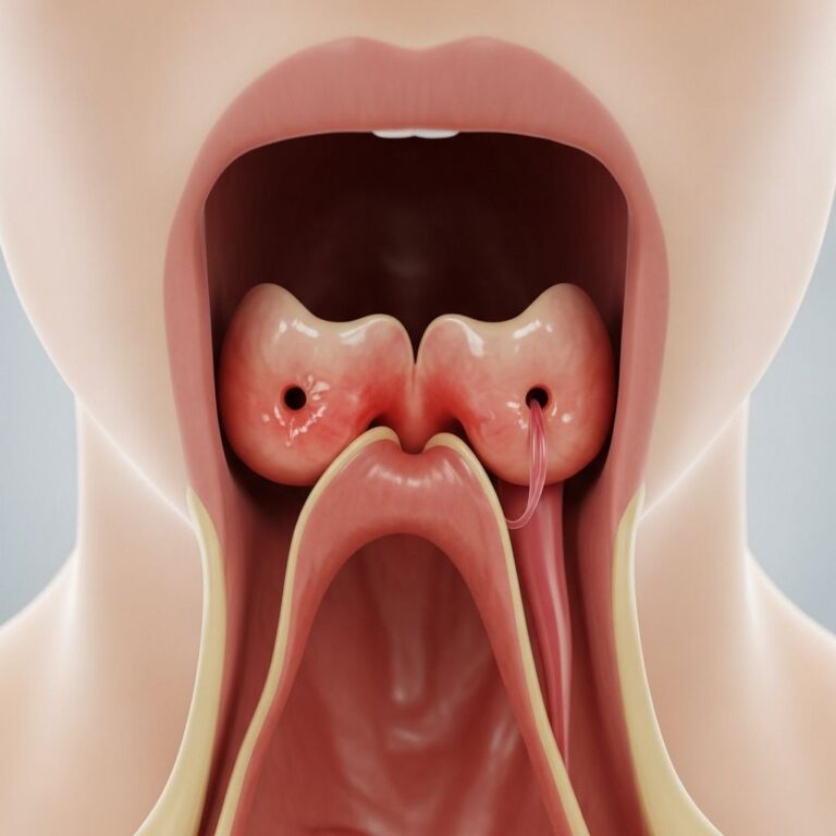
Xanthelasma, medically known as xanthelasma palpebrarum, is a benign skin condition characterized by harmless yellow plaques that appear on or near the eyelids. These deposits may present as soft, chalky, or semi-solid lesions and typically develop on the medial aspect of the upper eyelids, though they can also appear on the lower lids. The plaques are composed of cholesterol and other lipid deposits that accumulate in the skin over time. While xanthelasma is not inherently dangerous and does not have premalignant potential, it serves as an important visible marker of potential cholesterol imbalances and underlying metabolic conditions that warrant medical attention.
The condition is particularly significant because it often appears bilaterally and symmetrically above the inner corner of the eyes. Although the plaques themselves are painless and do not cause physical discomfort, many individuals seek treatment for cosmetic reasons, as the yellow appearance can affect self-confidence and appearance.
Causes and Risk Factors
Xanthelasma develops primarily due to elevated cholesterol levels and lipid metabolism disorders. The condition occurs when cholesterol and other lipid substances accumulate in the dermis layer of the skin around the eyelids. Several risk factors contribute to the development of xanthelasma:
- High cholesterol levels and dyslipidemia
- Genetic predisposition and familial hypercholesterolemia
- Metabolic disorders affecting lipid metabolism
- Liver disease and primary biliary cholangitis (PBC)
- Age-related changes in lipid processing
- Certain autoimmune and systemic conditions
The condition is closely associated with cardiovascular risk factors and can indicate an underlying metabolic imbalance that extends beyond cosmetic concerns. In some cases, xanthelasma may be one of several clinical manifestations of systemic diseases, making proper medical evaluation essential.
Recognizing Symptoms and Appearance
Xanthelasma presents with distinctive visual characteristics that make it relatively easy to identify. The primary symptom is the appearance of yellow or yellowish-orange plaques on the eyelids. These deposits typically manifest as:
- Bilateral yellow plaques on the medial upper eyelids
- Soft, chalky, or semi-solid texture
- Asymptomatic presentation (no pain or itching)
- Progressive enlargement over time
- Possible associated facial signs of high cholesterol
Many patients with xanthelasma also present with other cutaneous signs of high cholesterol, including arcus senilis (white or grayish rings around the cornea) and other xanthoma lesions on different body areas. The condition typically develops gradually and may remain stable or slowly enlarge over months to years.
Differential Diagnosis
While xanthelasma is generally straightforward to diagnose based on clinical presentation, several other conditions may resemble it and must be ruled out. These include necrobiotic xanthogranuloma, Erdheim-Chester disease, lipoid proteinosis, palpebral sarcoidosis, syringoma, sebaceous hyperplasia, and nodular basal cell carcinoma. Necrobiotic xanthogranuloma, a rare chronic granulomatous disease, presents with yellowish or violaceous plaques but is distinguished by its rarity, different etiology, and association with plasma cell dyscrasias. Patients presenting with atypical features or deviation from the classic bilateral medial canthal presentation should undergo more thorough evaluation to exclude these alternative diagnoses.
Diagnosis and Clinical Evaluation
The diagnosis of xanthelasma is typically clinical and based on characteristic appearance and patient history. A comprehensive evaluation includes:
- Physical examination of the eyelids and periorbital region
- Assessment of cholesterol levels through blood work
- Evaluation for associated systemic diseases
- Documentation of lesion size, location, and characteristics
- Screening for other cutaneous manifestations of dyslipidemia
While biopsy is not routinely necessary for diagnosis, it may be performed in ambiguous cases or when other conditions are suspected. Histopathological examination would reveal perivascular inflammatory infiltrate with mononucleated and multinucleated foamy histiocytes characterized by intracellular fat deposits, typically found in the upper reticular dermis.
Treatment Options
Cryotherapy Treatment
Cryotherapy is a widely accepted and effective method for xanthelasma removal, involving the application of extremely cold temperatures to eliminate the lipid deposits. The cryotherapy procedure typically follows these steps:
- Initial consultation to assess suitability for treatment
- Cleansing of the affected area to minimize infection risk
- Application of local anesthetic to reduce discomfort
- Careful application of liquid nitrogen using a cotton swab or spray
- Quick freezing of the xanthelasma deposit, lasting seconds to one minute
After treatment, the affected area develops a scab that heals over approximately two weeks. Common post-treatment effects include redness, swelling, and possible blistering, which are normal and typically subside naturally. Patients must avoid picking at the scab to prevent scarring and should protect the area from sunlight using appropriate sunscreen protection.
Laser Ablation
Laser ablation employs concentrated beams of light to vaporize xanthelasma with precision, reducing damage to surrounding tissues. This method is often preferred by dermatologists and oculoplastic surgeons due to its accuracy and minimal collateral tissue damage. Laser therapy allows for controlled removal of the lesion while preserving the delicate periocular skin architecture.
Electrodessication
Electrodessication utilizes electric currents to dry out xanthelasma, facilitating easier removal. This technique can be effective for smaller lesions and offers an alternative approach to liquid nitrogen or laser methods.
Chemical Peels
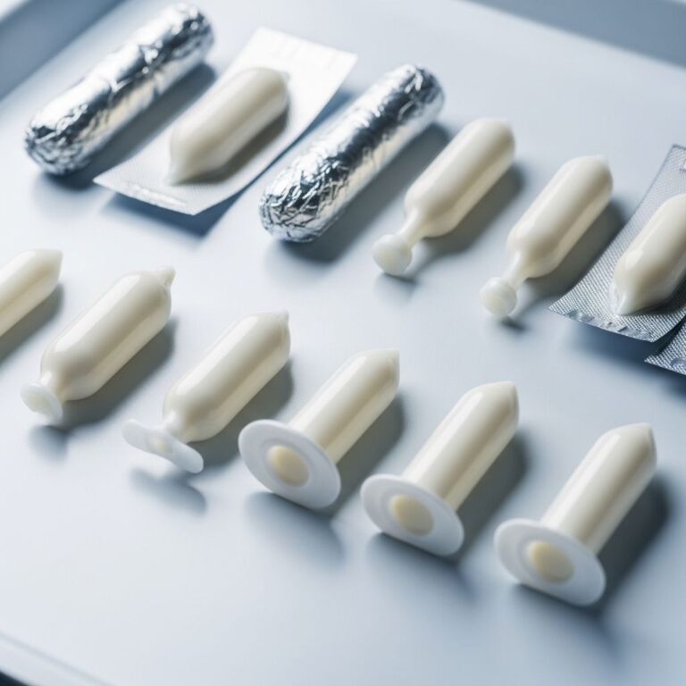
Chemical peels using bichloroacetic acid or trichloroacetic acid represent viable alternatives in xanthelasma management. Topical application of bichloroacetic acid offers advantages including simplicity and cost-effectiveness compared to other modalities. Radiofrequency treatments provide another option for lesion removal and skin rejuvenation.
Surgical Excision
For larger or more extensive xanthelasma lesions, surgical excision may be recommended. This approach involves direct removal of the affected tissue and may provide definitive results, though scarring risk must be considered based on lesion size and location.
Post-Treatment Care and Recovery
Successful xanthelasma treatment requires diligent post-treatment care to ensure optimal healing and minimize complications. Key aspects of aftercare include:
- Keeping the treated area clean and protected from contamination
- Applying prescribed antibacterial cream to support healing
- Avoiding sun exposure to the treated region
- Using appropriate sunscreen protection during healing
- Refraining from picking or disturbing the treated area
- Taking prescribed medications as directed for pain or infection prevention
- Monitoring for adverse reactions and reporting severe side effects
Once healed, the skin typically returns to its natural state with resolution of the yellow plaque appearance and restoration of normal eyelid contours.
Prevention and Recurrence Management
Even with successful treatment, xanthelasma can recur, making prevention strategies essential. Recurrence prevention focuses on managing underlying cholesterol levels and lipid metabolism through:
- Maintaining a heart-healthy diet low in saturated fats and cholesterol
- Engaging in regular physical exercise and maintaining healthy weight
- Taking cholesterol-lowering medications as prescribed
- Regular monitoring of lipid panel results
- Managing associated systemic conditions
- Attending follow-up appointments with healthcare providers
Collaboration with your primary care physician is essential to manage other risk factors that might contribute to recurrence. Chances of recurrence can be significantly reduced through comprehensive lipid management and appropriate lifestyle modifications.
Choosing the Right Treatment Provider
Selecting an appropriate healthcare provider significantly impacts treatment outcomes. The ideal provider should be a board-certified ophthalmologist or dermatologist with particular expertise in oculoplastic surgery and periocular conditions. Working with a specialist skilled in eyelid procedures enhances the likelihood of satisfactory cosmetic and functional outcomes while minimizing complications such as scarring or asymmetry.
Lifestyle Modifications for Prevention
Beyond direct treatment of xanthelasma, comprehensive lifestyle adjustments support prevention and reduce recurrence risk. These modifications include maintaining a balanced diet rich in fruits, vegetables, and whole grains while limiting saturated fats and refined sugars. Regular cardiovascular exercise, stress management, adequate sleep, and smoking cessation all contribute to improved lipid metabolism and overall health. Weight management and moderation of alcohol consumption further support metabolic health and cholesterol control.
Frequently Asked Questions
Q: Is xanthelasma dangerous or cancerous?
A: No, xanthelasma is a benign skin condition with no premalignant potential. However, it may indicate underlying cholesterol imbalances that warrant medical attention for cardiovascular health.
Q: Can xanthelasma be prevented?
A: While genetic factors play a role, maintaining healthy cholesterol levels through diet, exercise, and medication (if prescribed) can help prevent development or recurrence of xanthelasma.
Q: How long does xanthelasma treatment take?
A: The actual treatment procedure is quick, typically lasting seconds to one minute depending on lesion size. Complete healing usually requires two to four weeks.
Q: Will xanthelasma return after treatment?
A: Recurrence is common regardless of removal method, but the likelihood can be significantly reduced through cholesterol management and lifestyle modifications.
Q: Which treatment method is best for xanthelasma?
A: The optimal treatment depends on lesion size, severity, and individual factors. Laser therapy is often preferred, but cryotherapy, chemical peels, and surgical excision are viable alternatives that your doctor can recommend.
Q: Does xanthelasma cause pain or discomfort?
A: No, xanthelasma is typically asymptomatic and painless. Most patients seek treatment for cosmetic reasons rather than symptom relief.
References
- How To Get Rid Of Xanthelasma — Xanthelasma.com. Accessed 2025-12-01. https://xanthelasma.com/how-to-get-rid-of-xanthelasma/
- A Case Report of Xanthelasma and the Associated Differential Diagnosis — National Center for Biotechnology Information (NIH/NLM). PMC12064078. https://pmc.ncbi.nlm.nih.gov/articles/PMC12064078/
- Treatment of Xanthelasma Palpebrarum with Bichloracetic Acid — Journal of the American Academy of Dermatology. Wiley Online Library. https://onlinelibrary.wiley.com/doi/abs/10.1111/j.1524-4725.1998.tb04297.x
- High Cholesterol in Children: Symptoms, Causes & Treatment — Cleveland Clinic. 2024. https://my.clevelandclinic.org/health/diseases/12113-high-cholesterol-in-children
- Signs of High Cholesterol on the Face to Look For — Docus AI. Accessed 2025-12-01. https://docus.ai/symptoms-guide/high-cholesterol-signs-on-face
Similar Articles


Read full bio of Sneha Tete