Atopic Dermatitis Expert Guide: Causes, Symptoms & Treatment
Comprehensive guide to atopic dermatitis: causes, symptoms, diagnosis, and effective management strategies for all ages.

Author: Dermatological Expert
Revised: January 2026
What is atopic dermatitis?
Atopic dermatitis, commonly known as atopic eczema, is a chronic, relapsing inflammatory skin disorder characterised by dry skin and intensely itchy skin eruptions. It usually starts in early childhood, but can begin at any age, including during adulthood. The term ‘atopic’ implies a predisposition to allergic hypersensitivity reactions such as allergic rhinitis, asthma and conjunctivitis.
Atopic dermatitis affects 15–30% of children and 2–10% of adults worldwide. It is more common in higher socioeconomic groups and urban areas. There is often a family history of atopic dermatitis, allergic rhinitis or asthma. The condition is more common in people who have a personal or family history of allergic conditions such as allergic rhinitis, asthma or allergic conjunctivitis (atopic triad).
The management of atopic dermatitis should be individualised based on severity, considering patient preferences, costs, and adverse effects. General measures include trigger avoidance, skin hydration with moisturisers, education, pruritus control, and acute flare management.
Who gets atopic dermatitis?
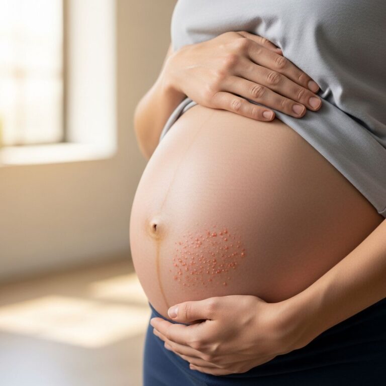
Atopic dermatitis can affect people of any age, race, sex or geographic location. However, it most often appears in early childhood and can persist into adulthood. Up to 20% of children are affected, with 60% developing symptoms in their first year of life and 85% before age 5. About half of affected children will continue to have symptoms into adulthood.
- Infants: often on cheeks, scalp and limbs
- Children: flexural areas (antecubital and popliteal fossae)
- Adults: hands, eyelids, neck, and flexural areas
Risk factors include family history of atopy, urban environment, and higher socioeconomic status.
What causes atopic dermatitis?
The exact cause of atopic dermatitis is unknown. It arises from a complex interaction of genetic, immunological, environmental and skin barrier factors. Multiple genes are involved, with mutations in filaggrin (FLG) gene being the strongest predisposing factor, leading to impaired skin barrier function.
Key pathogenic mechanisms include:
- Immune dysregulation: Th2-dominated response with elevated IgE, IL-4, IL-13, IL-31
- Skin barrier defects: Reduced ceramides, filaggrin mutations causing xerosis
- Microbiome dysbiosis: Staphylococcus aureus overgrowth
- Pruritus-scratch cycle: Itch leads to scratching, worsening barrier damage
Environmental triggers exacerbate the genetic predisposition, including irritants, allergens, climate changes, and infections.
What are the clinical features of atopic dermatitis?
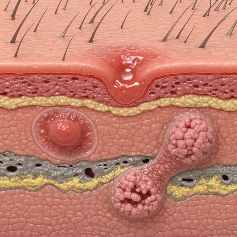
Atopic dermatitis has a highly variable clinical course with acute, subacute, and chronic phases. The cardinal features are pruritus and xerosis, with characteristic morphology and distribution varying by age.
Infantile phase (2 months–2 years)
- Acute: weepy, crusted, erythematous papules and plaques on cheeks, scalp, trunk
- Predilection for extensor surfaces
Childhood phase (2–puberty)
- Lichenification and scaling in flexural areas (antecubital/popliteal fossae)
- Face, neck, wrists, ankles involved
Adult phase
- Thickened lichenified plaques in flexural areas, hands, eyelids
- Head and neck dermatitis common
Diagnostic criteria (UK Working Party):
| Major features (≥3 required) |
|---|
| Must have itchy skin |
| Typical morphology and distribution |
| Chronic/relapsing course |
| Personal/family atopy |
| Minor features (≥3) |
|---|
| Xerosis, ichthyosis, hyperlinear palms |
| Keratoconus, nipple eczema |
| Dennie-Morgan folds, pityriasis alba |
Severity assessment tools: SCORAD, EASI, POEM.
Diagnosis
Diagnosis is primarily clinical based on history and examination. No single test confirms atopic dermatitis; it relies on characteristic features and exclusion of mimics. Patch testing may identify contact allergens. Elevated IgE and eosinophilia support diagnosis but are not specific.
- Biopsy rarely needed but shows spongiosis, acanthosis, lymphocytic infiltrate
- Skin swabs for secondary infection (S. aureus, HSV)
Complications
Atopic dermatitis predisposes to numerous complications due to impaired barrier and immune dysregulation.
Bacterial infections
Staphylococcus aureus colonisation in 90% of patients, leading to impetigo, folliculitis, cellulitis. Treat with topical antiseptics, antibiotics for overt infection.
Viral infections
- Herpes simplex (eczema herpeticum): Monomorphic punched-out erosions; systemic aciclovir required
- Molluscum contagiosum, warts more severe
Fungal infections
Malassezia in head/neck dermatitis; topical antifungals effective.
Contact dermatitis
Irritant (soaps, wool) or allergic (preservatives, fragrances).
Psychosocial impact
Sleep disturbance, anxiety, depression, stigmatisation.
Ocular complications
Keratoconus, cataracts, ocular rosacea.
Hand dermatitis
Common in adults, occupational triggers.
Treatment of atopic dermatitis
Treatment targets inflammation control, barrier repair, itch reduction, and infection prevention. A stepwise approach from mild topical therapy to systemic agents for severe/refractory disease.
Basic skin care
- Daily emollient use: Apply liberally 2–3 times daily, especially after bathing
- Soap substitutes, short lukewarm baths (5–10 min)
- Bleach baths (½ cup per full tub) twice weekly for S. aureus
Topical anti-inflammatory therapy
| Agent | Class | Use |
|---|---|---|
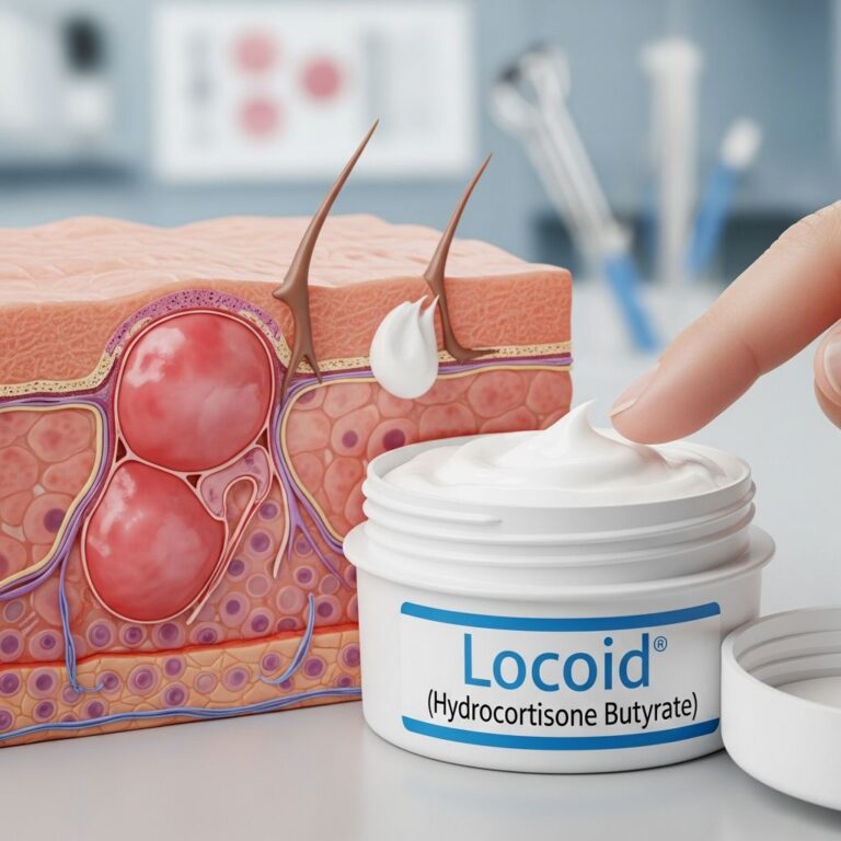
| Topical corticosteroids (TCS) | Mild-moderate potency | First-line for flares; proactive twice weekly |
| TCIs (tacrolimus, pimecrolimus) | Non-steroidal | Face, maintenance, steroid-sparing |
| PDE4 inhibitor (crisaborole) | Mild-moderate | Children ≥2 years |
| JAK inhibitors (ruxolitinib cream) | Targeted | Non-responsive areas |
Systemic therapy (moderate-severe)
- Phototherapy: NB-UVB first-line
- Biologics: Dupilumab (anti-IL4/13), tralokinumab
- JAK inhibitors: Upadacitinib, abrocitinib oral
- Immunosuppressants: Cyclosporine, methotrexate
Wet wrap therapy
For severe flares: TCS + damp wraps + dry layer overnight.
Allergen/immunotherapy
Avoidance of identified triggers; allergen immunotherapy for house dust mite in selected cases.
Prevention and prognosis
Early emollient use may prevent atopic dermatitis in high-risk infants. Prognosis improves with age; 60–70% remit by adolescence, but relapses common. Continued moisturiser use prevents flares.
Frequently Asked Questions
What is the best moisturiser for atopic dermatitis?
Thick, ointment-based emollients with ceramides, applied frequently. Patient preference important.
Can atopic dermatitis be cured?
No cure, but can be well-controlled with consistent treatment.
Is bleach bath safe for children?
Yes, dilute bleach baths 2x/week reduce S. aureus.
When to use steroid creams?
Twice daily during flares until clear, then proactive maintenance.
What if topical treatments fail?
Escalate to phototherapy, biologics, or systemic JAK inhibitors.
References
- Atopic Dermatitis: A Review of Diagnosis and Treatment — PMC / National Library of Medicine. 2024. https://pmc.ncbi.nlm.nih.gov/articles/PMC11627575/
- Atopic Dermatitis – A Guide for Your Family — Boston Children’s Hospital. 2025-02. https://www.childrenshospital.org/sites/default/files/2025-02/atopic-dermatitis-guide-english.pdf
- Atopic dermatitis clinical guideline — American Academy of Dermatology (AAD). 2025. https://www.aad.org/member/clinical-quality/guidelines/atopic-dermatitis
- Atopic Dermatitis: A Practical Guide to Management — Skin Therapy Letter. Recent. https://www.skintherapyletter.com/atopic-dermatitis/management-guide/
- Treatment of Atopic Dermatitis — American Academy of Pediatrics (AAP). Recent. https://www.aap.org/en/patient-care/atopic-dermatitis/treatment-of-atopic-dermatitis/
- Atopic dermatitis (eczema) – Diagnosis and treatment — Mayo Clinic. Recent. https://www.mayoclinic.org/diseases-conditions/atopic-dermatitis-eczema/diagnosis-treatment/drc-20353279
Similar Articles


Read full bio of Sneha Tete