Epidermoid Cyst: Key Insights On Symptoms, Causes & Management
Comprehensive guide to epidermoid cysts: causes, symptoms, diagnosis, treatment, and prevention strategies for skin health.

Epidermoid cysts, often misnamed as sebaceous cysts, represent the most prevalent type of cutaneous cysts. These benign, encapsulated subepidermal nodules contain keratin material derived from the epidermis. Typically appearing on the face, neck, trunk, or genitals, they form due to proliferation of surface epidermal cells within the dermis. While generally harmless, they can become inflamed, infected, or rarely malignant, necessitating awareness for proper management.
What is an epidermoid cyst?
An
epidermoid cyst
is a slow-growing, non-cancerous lump beneath the skin’s surface, filled with keratin—a thick, protein-rich substance produced by skin cells. Unlike true sebaceous cysts, which involve sebaceous glands, epidermoid cysts originate from epidermal cells trapped under the skin. They feature a central punctum (blackhead-like opening) through which cheesy, foul-smelling material may drain if ruptured. These cysts are firm, mobile, and dome-shaped, ranging from 0.5 to 5 cm in diameter, though larger ones occur rarely.Histologically, the cyst wall consists of stratified squamous epithelium resembling the epidermis, continuously shedding keratin into the cystic cavity. This accumulation causes gradual expansion. In newborns, similar tiny cysts called milia are common and often resolve spontaneously.
Who gets epidermoid cysts?
Epidermoid cysts predominantly affect adults in their third and fourth decades, with a male-to-female ratio of 2:1. They are rare before puberty but milia-like cysts appear in up to 40-50% of newborns. Prevalence increases with age, particularly in sun-exposed areas due to cumulative damage. Genetic predispositions occur in syndromes like Gardner syndrome, where multiple cysts accompany colon polyps.
- **Common sites:** Face (cheeks, forehead), neck, trunk, scalp, ears, genitals, and extremities.
- **Risk factors:** Acne vulgaris, skin trauma, chronic sun exposure, medications (e.g., BRAF inhibitors, cyclosporine), and HPV infection.
- **Rare associations:** Favre-Racouchot syndrome (solar elastosis with cysts in elderly), Gardner syndrome.
What causes epidermoid cysts?
The primary mechanism involves entrapment of epidermal cells beneath the skin surface, leading to cyst formation. Common triggers include:
- Blockage of hair follicle infundibulum, causing keratin buildup.
- Skin trauma or surgery implanting epidermal cells into dermis.
- Developmental anomalies, as in milia.
- Chronic UV exposure promoting cyst development in actinically damaged skin.
- Drug-induced: BRAF inhibitors, imiquimod, cyclosporine.
Pathophysiology centers on follicular orifice plugging, with the cyst communicating via a keratin-filled punctum. Rupture displaces keratin into dermis, provoking intense inflammation via foreign body reaction.
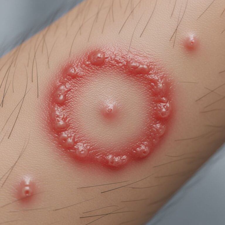
What are the clinical features of epidermoid cyst?
Most cysts are asymptomatic, presenting as smooth, round, firm nodules with a central blackhead. Skin overlying is normal unless inflamed. Sizes vary from pea-sized to golf ball-sized.
| Stage | Features |
|---|---|
| Uncomplicated | Firm, mobile, painless nodule; central punctum; cheesy discharge if expressed. |
| Inflamed | Tender, red, swollen; foul-smelling pus if infected. |
| Ruptured | Sudden pain, boggy swelling, granulomatous reaction. |
| Infected | Abscess formation, fever, lymphangitis. |
Rare complications: Malignant transformation to squamous cell carcinoma (1% risk), especially in longstanding or traumatized cysts.
Diagnosis
Diagnosis relies on clinical examination: characteristic nodule with punctum suffices in most cases. No routine labs or imaging needed. Incisional biopsy if atypical (rapid growth, irregular borders, ulceration). Dermoscopy reveals translucent keratin plug. Ultrasound confirms cystic nature if deep.
- **When to biopsy:** Painful, growing, recurrent, or in high-risk sites (digits, palms).
- **Histology:** Cyst lined by squamous epithelium with keratin flakes; inflammation if ruptured.
Differential diagnosis
Key mimics include:
- **Pilar (trichilemmal) cyst:** Scalp predilection, lacks punctum, family history.
- **Lipoma:** Softer, no punctum.
- **Furuncle/carbuncle:** Acute inflammation, resolves with antibiotics.
- **Dermoid cyst:** Congenital, midline.
- **Steatocystoma multiplex:** Multiple, translucent.
- **Branchial cleft cyst:** Neck, fluctuant.
- **Pilonidal cyst:** Sacrococcygeal.
- **Calcinosis cutis:** Hard, calcium deposits.
Management
Asymptomatic cysts require no treatment—reassurance suffices. Intervention for cosmetics, symptoms, or complications.
Conservative
- Warm compresses for inflammation.
- Intralesional steroids (triamcinolone 10-40 mg/mL) for inflamed cysts.
- Antibiotics if infected.
Surgical
Gold standard: Complete excision with intact cyst wall under local anesthesia. Delay if acutely inflamed; perform incision-drainage first.
- **Minimal excision:** Punch biopsy tool extracts cyst via small hole.
- **Incision + curettage:** For ruptured cysts.
- **Post-op:** Minimal scarring, low recurrence (<5%) if wall fully removed.
Avoid simple drainage—recurrence nears 50% without wall excision.
Prevention
- Avoid skin trauma and picking acne.
- Sun protection to minimize UV-induced cysts.
- Early acne treatment reduces comedone-derived cysts.
- Monitor high-risk patients (Gardner syndrome).
Complications
- Inflammation/infection (most common).
- Recurrence post-incomplete removal.
- Scarring from rupture or surgery.
- Rare malignancy (SCC/BCC).
Frequently Asked Questions (FAQs)
Q: Do epidermoid cysts go away on their own?
A: Rarely; they persist and may enlarge unless excised. Inflamed ones may resolve temporarily but recur.
Q: Are epidermoid cysts cancerous?
A: Benign in >99%; rare transformation to SCC, especially if traumatized repeatedly.
Q: Can I squeeze an epidermoid cyst?
A: No—risks inflammation, infection, and scarring. Seek professional removal.
Q: How much does cyst removal cost?
A: Varies; often covered if symptomatic. Outpatient procedure minimizes expense.
Q: Will removal leave a scar?
A: Minimal scar with proper technique; fades over months.
Q: Can cysts appear on fingers or toes?
A: Yes, rare but prompts Gardner syndrome evaluation.
References
- Epidermoid Cyst – StatPearls — NCBI Bookshelf. 2023-07-17. https://www.ncbi.nlm.nih.gov/books/NBK499974/
- Epidermoid Cyst Signs, Symptoms, Removal & Surgery — Pacific Neuroscience Institute. 2024-01-15. https://www.pacificneuroscienceinstitute.org/brain-tumor/conditions/epidermoid-cyst/
- Epidermoid Cysts of the Skin — UR Medicine / University of Rochester. 2023-11-01. https://www.urmc.rochester.edu/encyclopedia/content?contenttypeid=85&contentid=P03392
- Epidermal Inclusion Cysts (Sebaceous Cysts): Treatment & Causes — Cleveland Clinic. 2024-05-20. https://my.clevelandclinic.org/health/diseases/14165-sebaceous-cysts
- Epidermoid cyst — MedlinePlus / NIH. 2023-09-12. https://medlineplus.gov/ency/article/000842.htm
- Epidermoid cysts — UM Health-Sparrow. 2024-02-28. https://www.uofmhealthsparrow.org/departments-conditions-conditions/epidermoid-cysts
- Epidermoid cysts – Symptoms and causes — Mayo Clinic. 2024-06-10. https://www.mayoclinic.org/diseases-conditions/epidermoid-cysts/symptoms-causes/syc-20352701
Similar Articles


Read full bio of Sneha Tete